 Movie
Movie Controller


+ Open data

- Basic information
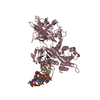
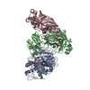
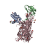
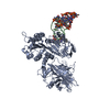
| Entry | Database: PDB / ID: 1gji | ||||||
|---|---|---|---|---|---|---|---|
| Title | Crystal structure of c-Rel bound to DNA | ||||||
 Components Components |
| ||||||
 Keywords Keywords | TRANSCRIPTION/DNA / NF-kB-DNA COMPLEX / TRANSCRIPTION FACTOR / c-Rel HOMODIMER / TRANSCRIPTION-DNA COMPLEX | ||||||
| Function / homology |  Function and homology information Function and homology informationapoptotic DNA fragmentation / non-canonical NF-kappaB signal transduction / canonical NF-kappaB signal transduction / response to cytokine / DNA-binding transcription factor activity, RNA polymerase II-specific / RNA polymerase II cis-regulatory region sequence-specific DNA binding / DNA-binding transcription factor activity / inflammatory response / innate immune response / positive regulation of transcription by RNA polymerase II ...apoptotic DNA fragmentation / non-canonical NF-kappaB signal transduction / canonical NF-kappaB signal transduction / response to cytokine / DNA-binding transcription factor activity, RNA polymerase II-specific / RNA polymerase II cis-regulatory region sequence-specific DNA binding / DNA-binding transcription factor activity / inflammatory response / innate immune response / positive regulation of transcription by RNA polymerase II / DNA binding / nucleus / cytoplasm Similarity search - Function | ||||||
| Biological species |  | ||||||
| Method |  X-RAY DIFFRACTION / molecular replacement, MIR / Resolution: 2.85 Å X-RAY DIFFRACTION / molecular replacement, MIR / Resolution: 2.85 Å | ||||||
 Authors Authors | Huang, D.B. / Chen, Y.Q. / Ruetsche, M. / Phelps, C.B. / Ghosh, G. | ||||||
 Citation Citation | Journal: Structure / Year: 2001 Title: X-ray crystal structure of proto-oncogene product c-Rel bound to the CD28 response element of IL-2. Authors: Huang, D.B. / Chen, Y.Q. / Ruetsche, M. / Phelps, C.B. / Ghosh, G. | ||||||
| History |
|
- Structure visualization
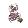
| Structure viewer | Molecule: MolmilJmol/JSmol |
|---|
- Downloads & links
-Download
| PDBx/mmCIF format | 1gji.cif.gz | 159 KB | Display | PDBx/mmCIF format |
|---|---|---|---|---|
| PDB format | pdb1gji.ent.gz | 127.1 KB | Display | PDB format |
| PDBx/mmJSON format | 1gji.json.gz | Tree view | PDBx/mmJSON format | |
| Others |  Other downloads Other downloads |
-Validation report
| Arichive directory | https://data.pdbj.org/pub/pdb/validation_reports/gj/1gjiftp://data.pdbj.org/pub/pdb/validation_reports/gj/1gji | HTTPS FTP |
|---|
-Related structure data
| Similar structure data |
|---|

-Links
 PDBj
PDBj


- Assembly
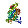
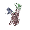
| Deposited unit | 
| ||||||||
|---|---|---|---|---|---|---|---|---|---|
| 1 |
| ||||||||
| Unit cell |
|
-Components
| #1: DNA chain | Mass: 6206.054 Da / Num. of mol.: 1 / Source method: obtained synthetically |
|---|---|
| #2: DNA chain | Mass: 6058.939 Da / Num. of mol.: 1 / Source method: obtained synthetically |
| #3: Protein | Mass: 31894.316 Da / Num. of mol.: 2 / Fragment: Rel homology region Source method: isolated from a genetically manipulated source Source: (gene. exp.)  |
-Experimental details
-Experiment
| Experiment | Method: X-RAY DIFFRACTION / Number of used crystals: 1 |
|---|
- Sample preparation
| Crystal | Density Matthews: 3.16 Å3/Da / Density % sol: 63 % | ||||||||||||||||||||||||||||||||||||||||||
|---|---|---|---|---|---|---|---|---|---|---|---|---|---|---|---|---|---|---|---|---|---|---|---|---|---|---|---|---|---|---|---|---|---|---|---|---|---|---|---|---|---|---|---|
| Crystal grow | Temperature: 298 K / Method: evaporation / pH: 7.5 Details: PEG 4000, spermine, DTT, pH 7.5, EVAPORATION, temperature 298.0K | ||||||||||||||||||||||||||||||||||||||||||
| Components of the solutions |
| ||||||||||||||||||||||||||||||||||||||||||
| Crystal grow | *PLUS Method: vapor diffusion, hanging drop | ||||||||||||||||||||||||||||||||||||||||||
| Components of the solutions | *PLUS
|
-Data collection
| Diffraction | Mean temperature: 168 K |
|---|---|
| Diffraction source | Source: ROTATING ANODE / Type: RIGAKU RU200 / Wavelength: 1.5418 |
| Detector | Type: MARRESEARCH / Detector: AREA DETECTOR / Date: Feb 14, 1999 |
| Radiation | Protocol: SINGLE WAVELENGTH / Monochromatic (M) / Laue (L): M / Scattering type: x-ray |
| Radiation wavelength | Wavelength: 1.5418 Å / Relative weight: 1 |
| Reflection | Resolution: 2.85→25 Å / Num. all: 21455 / Num. obs: 18723 / % possible obs: 90.8 % / Observed criterion σ(F): 2 / Redundancy: 10 % / Rmerge(I) obs: 0.086 / Net I/σ(I): 7.8 |
| Reflection shell | Resolution: 2.85→2.95 Å / Redundancy: 7 % / Rmerge(I) obs: 0.424 / Num. unique all: 1159 / % possible all: 46 |
| Reflection | *PLUS Num. obs: 21455 / % possible obs: 90 % / Num. measured all: 215187 / Rmerge(I) obs: 0.086 |
| Reflection shell | *PLUS % possible obs: 48 % / Rmerge(I) obs: 0.424 / Mean I/σ(I) obs: 1.8 |
- Processing
| Software |
| ||||||||||||||||||||
|---|---|---|---|---|---|---|---|---|---|---|---|---|---|---|---|---|---|---|---|---|---|
| Refinement | Method to determine structure: molecular replacement, MIR Starting model: p65/DNA complex Resolution: 2.85→25 Å / σ(F): 2 / Stereochemistry target values: Engh & Huber Details: The DNA molecule has alternate conformations. Two sets of coordinates for the DNA molecule were refined with 0.5 occupancy each.
| ||||||||||||||||||||
| Refinement step | Cycle: LAST / Resolution: 2.85→25 Å
| ||||||||||||||||||||
| Refine LS restraints |
| ||||||||||||||||||||
| Refinement | *PLUS % reflection Rfree: 5 % / Rfactor obs: 0.227 / Rfactor Rfree: 0.279 / Rfactor Rwork: 0.227 | ||||||||||||||||||||
| Solvent computation | *PLUS | ||||||||||||||||||||
| Displacement parameters | *PLUS | ||||||||||||||||||||
| Refine LS restraints | *PLUS Type: c_bond_d / Dev ideal: 0.0079 |
