[English] 日本語
 Yorodumi
- PDB-1ghj: SOLUTION STRUCTURE OF THE LIPOYL DOMAIN OF THE 2-OXOGLUTARATE DEH... -
+ Open data
- Basic information
| Entry | Database: PDB / ID: 1ghj | ||||||
|---|---|---|---|---|---|---|---|
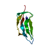
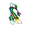
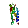
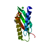
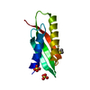
| Title | SOLUTION STRUCTURE OF THE LIPOYL DOMAIN OF THE 2-OXOGLUTARATE DEHYDROGENASE COMPLEX FROM AZOTOBACTER VINELAND II, NMR, MINIMIZED AVERAGE STRUCTURE | ||||||
 Components Components | E2, THE DIHYDROLIPOAMIDE SUCCINYLTRANSFERASE COMPONENT OF 2-OXOGLUTARATE DEHYDROGENASE COMPLEX | ||||||
 Keywords Keywords | ACYLTRANSFERASE / GLYCOLYSIS / TRANSFERASE / LIPOYL | ||||||
| Function / homology |  Function and homology information Function and homology informationL-lysine catabolic process to acetyl-CoA via saccharopine / dihydrolipoyllysine-residue succinyltransferase / dihydrolipoyllysine-residue succinyltransferase activity / oxoglutarate dehydrogenase complex / tricarboxylic acid cycle / cytosol Similarity search - Function | ||||||
| Biological species |  Azotobacter vinelandii (bacteria) Azotobacter vinelandii (bacteria) | ||||||
| Method | SOLUTION NMR | ||||||
 Authors Authors | Berg, A. / Vervoort, J. / De Kok, A. | ||||||
 Citation Citation |  Journal: J.Mol.Biol. / Year: 1996 Journal: J.Mol.Biol. / Year: 1996Title: Solution structure of the lipoyl domain of the 2-oxoglutarate dehydrogenase complex from Azotobacter vinelandii. Authors: Berg, A. / Vervoort, J. / de Kok, A. #1:  Journal: Eur.J.Biochem. / Year: 1995 Journal: Eur.J.Biochem. / Year: 1995Title: Sequential 1H and 15N Nuclear Magnetic Resonance Assignments and Secondary Structure of the Lipoyl Domain of the 2-Oxoglutarate Dehydrogenase Complex from Azotobacter Vinelandii. Evidence for ...Title: Sequential 1H and 15N Nuclear Magnetic Resonance Assignments and Secondary Structure of the Lipoyl Domain of the 2-Oxoglutarate Dehydrogenase Complex from Azotobacter Vinelandii. Evidence for High Structural Similarity with the Lipoyl Domain of the Pyruvate Dehydrogenase Complex Authors: Berg, A. / Smits, O. / De Kok, A. / Vervoort, J. | ||||||
| History |
|
- Structure visualization
| Structure viewer | Molecule:  Molmil Molmil Jmol/JSmol Jmol/JSmol |
|---|
- Downloads & links
- Download
| PDBx/mmCIF format |  1ghj.cif.gz 1ghj.cif.gz | 35.1 KB | Display |  PDBx/mmCIF format PDBx/mmCIF format |
|---|---|---|---|---|
| PDB format |  pdb1ghj.ent.gz pdb1ghj.ent.gz | 24.7 KB | Display |  PDB format PDB format |
| PDBx/mmJSON format |  1ghj.json.gz 1ghj.json.gz | Tree view |  PDBx/mmJSON format PDBx/mmJSON format | |
| Others |  Other downloads Other downloads |
-Validation report
| Summary document |  1ghj_validation.pdf.gz 1ghj_validation.pdf.gz | 333.4 KB | Display |  wwPDB validaton report wwPDB validaton report |
|---|---|---|---|---|
| Full document |  1ghj_full_validation.pdf.gz 1ghj_full_validation.pdf.gz | 336.1 KB | Display | |
| Data in XML |  1ghj_validation.xml.gz 1ghj_validation.xml.gz | 3.4 KB | Display | |
| Data in CIF |  1ghj_validation.cif.gz 1ghj_validation.cif.gz | 4.1 KB | Display | |
| Arichive directory |  https://data.pdbj.org/pub/pdb/validation_reports/gh/1ghj https://data.pdbj.org/pub/pdb/validation_reports/gh/1ghj ftp://data.pdbj.org/pub/pdb/validation_reports/gh/1ghj ftp://data.pdbj.org/pub/pdb/validation_reports/gh/1ghj | HTTPS FTP |
-Related structure data
- Links
Links
- Assembly
| Deposited unit | 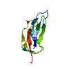
| |||||||||
|---|---|---|---|---|---|---|---|---|---|---|
| 1 |
| |||||||||
| NMR ensembles |
|
- Components
| #1: Protein | Mass: 8353.437 Da / Num. of mol.: 1 / Fragment: LIPOYL DOMAIN, RESIDUES 1-79 Source method: isolated from a genetically manipulated source Source: (gene. exp.)  Azotobacter vinelandii (bacteria) / Production host: Azotobacter vinelandii (bacteria) / Production host:  References: UniProt: P20708, dihydrolipoyllysine-residue succinyltransferase |
|---|
-Experimental details
-Experiment
| Experiment | Method: SOLUTION NMR |
|---|
- Sample preparation
| Crystal grow | *PLUS Method: other / Details: NMR |
|---|
- Processing
| Software |
| ||||||||||||
|---|---|---|---|---|---|---|---|---|---|---|---|---|---|
| NMR software | Name:  X-PLOR / Version: 3.1 / Developer: BRUNGER / Classification: refinement X-PLOR / Version: 3.1 / Developer: BRUNGER / Classification: refinement | ||||||||||||
| NMR ensemble | Conformers submitted total number: 1 |
 Movie
Movie Controller







 PDBj
PDBj

