 Movie
Movie Controller

[English] 日本語
 Yorodumi
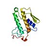
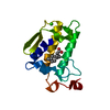
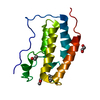
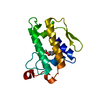
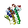
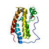
- PDB-1gh4: Structure of the triple mutant (K56M, K120M, K121M) of phospholip... -
+ Open data

- Basic information
| Entry | Database: PDB / ID: 1gh4 | ||||||
|---|---|---|---|---|---|---|---|
| Title | Structure of the triple mutant (K56M, K120M, K121M) of phospholipase A2 | ||||||
 Components Components | PHOSPHOLIPASE A2 | ||||||
 Keywords Keywords | HYDROLASE / Alpha Helix / Beta Sheet / Triple mutant | ||||||
| Function / homology |  Function and homology information Function and homology informationAcyl chain remodelling of PS / Acyl chain remodelling of PG / Synthesis of PA / Acyl chain remodelling of PC / Acyl chain remodelling of PE / Acyl chain remodelling of PI / positive regulation of podocyte apoptotic process / phosphatidylglycerol metabolic process / phosphatidylcholine metabolic process / bile acid binding ...Acyl chain remodelling of PS / Acyl chain remodelling of PG / Synthesis of PA / Acyl chain remodelling of PC / Acyl chain remodelling of PE / Acyl chain remodelling of PI / positive regulation of podocyte apoptotic process / phosphatidylglycerol metabolic process / phosphatidylcholine metabolic process / bile acid binding / phospholipase A2 / : / arachidonate secretion / lipid catabolic process / innate immune response in mucosa / phospholipid binding / positive regulation of fibroblast proliferation / fatty acid biosynthetic process / antimicrobial humoral immune response mediated by antimicrobial peptide / antibacterial humoral response / defense response to Gram-positive bacterium / signaling receptor binding / calcium ion binding / cell surface / : Similarity search - Function | ||||||
| Biological species |  | ||||||
| Method |  X-RAY DIFFRACTION / Resolution: 1.9 Å X-RAY DIFFRACTION / Resolution: 1.9 Å | ||||||
 Authors Authors | Sekar, K. / Velmurugan, D. / Tsai, M.D. | ||||||
 Citation Citation | Journal: J.Mol.Biol. / Year: 2002 Title: Observation of additional calcium ion in the crystal structure of the triple mutant K56,120,121M of bovine pancreatic phospholipase A2. Authors: Rajakannan, V. / Yogavel, M. / Poi, M.J. / Jeyaprakash, A.A. / Jeyakanthan, J. / Velmurugan, D. / Tsai, M.D. / Sekar, K. | ||||||
| History |
|
- Structure visualization
| Structure viewer | Molecule: MolmilJmol/JSmol |
|---|
- Downloads & links
-Download
| PDBx/mmCIF format | 1gh4.cif.gz | 41 KB | Display | PDBx/mmCIF format |
|---|---|---|---|---|
| PDB format | pdb1gh4.ent.gz | 27.6 KB | Display | PDB format |
| PDBx/mmJSON format | 1gh4.json.gz | Tree view | PDBx/mmJSON format | |
| Others |  Other downloads Other downloads |
-Validation report
| Arichive directory | https://data.pdbj.org/pub/pdb/validation_reports/gh/1gh4ftp://data.pdbj.org/pub/pdb/validation_reports/gh/1gh4 | HTTPS FTP |
|---|
-Related structure data
| Related structure data | |
|---|---|
| Similar structure data |


-Links
 PDBj
PDBj

- Assembly
| Deposited unit | 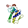
| ||||||||
|---|---|---|---|---|---|---|---|---|---|
| 1 |
| ||||||||
| Unit cell |
|
-Components
| #1: Protein | Mass: 13816.551 Da / Num. of mol.: 1 / Mutation: K56M,K120M,K121M Source method: isolated from a genetically manipulated source Source: (gene. exp.)  | ||||||
|---|---|---|---|---|---|---|---|
| #2: Chemical |   Mass: 40.078 Da / Num. of mol.: 2 / Source method: obtained synthetically / Formula: Ca Mass: 40.078 Da / Num. of mol.: 2 / Source method: obtained synthetically / Formula: Ca#3: Chemical | ChemComp-MPD / ( |   Mass: 118.174 Da / Num. of mol.: 1 / Source method: obtained synthetically / Formula: C6H14O2 / Comment: precipitant*YM Mass: 118.174 Da / Num. of mol.: 1 / Source method: obtained synthetically / Formula: C6H14O2 / Comment: precipitant*YM#4: Water | ChemComp-HOH / |  Mass: 18.015 Da / Num. of mol.: 125 / Source method: isolated from a natural source / Formula: H2O Mass: 18.015 Da / Num. of mol.: 125 / Source method: isolated from a natural source / Formula: H2OHas protein modification | Y | |
-Experimental details
-Experiment
| Experiment | Method: X-RAY DIFFRACTION / Number of used crystals: 1 |
|---|
- Sample preparation
| Crystal | Density Matthews: 2.26 Å3/Da / Density % sol: 45.59 % | |||||||||||||||||||||||||
|---|---|---|---|---|---|---|---|---|---|---|---|---|---|---|---|---|---|---|---|---|---|---|---|---|---|---|
| Crystal grow | Temperature: 293 K / Method: vapor diffusion method / pH: 7.2 Details: Tris Buffer, MPD, 70% MPD reservoir, 17-20Mg/ml protein, 5 mM CaCl2, pH 7.2, vapor diffusion method, temperature 293.0K | |||||||||||||||||||||||||
| Crystal grow | *PLUS Method: vapor diffusion, hanging drop | |||||||||||||||||||||||||
| Components of the solutions | *PLUS
|
-Data collection
| Diffraction | Mean temperature: 293 K |
|---|---|
| Diffraction source | Source: ROTATING ANODE / Wavelength: 1.5418 |
| Detector | Type: MARRESEARCH / Detector: IMAGE PLATE / Date: Aug 20, 2000 |
| Radiation | Protocol: SINGLE WAVELENGTH / Monochromatic (M) / Laue (L): M / Scattering type: x-ray |
| Radiation wavelength | Wavelength: 1.5418 Å / Relative weight: 1 |
| Reflection | Resolution: 1.9→14.2 Å / Num. all: 8880 / Num. obs: 8880 / % possible obs: 88 % / Redundancy: 4.6 % / Rmerge(I) obs: 0.075 |
| Reflection shell | Resolution: 1.9→1.97 Å / Rmerge(I) obs: 0.293 / Num. unique all: 910 / % possible all: 72 |
| Reflection | *PLUS % possible obs: 88.3 % / Num. measured all: 40817 |
- Processing
| Software |
| ||||||||||||||||||||||||
|---|---|---|---|---|---|---|---|---|---|---|---|---|---|---|---|---|---|---|---|---|---|---|---|---|---|
| Refinement | Resolution: 1.9→14.2 Å / σ(F): 0 / σ(I): 0
| ||||||||||||||||||||||||
| Refinement step | Cycle: LAST / Resolution: 1.9→14.2 Å
| ||||||||||||||||||||||||
| Refine LS restraints | Type: protein_rep.param / Dev ideal: 0.011 | ||||||||||||||||||||||||
| Software | *PLUS Name: CNS / Version: 1 / Classification: refinement | ||||||||||||||||||||||||
| Refinement | *PLUS Highest resolution: 1.9 Å / Lowest resolution: 14.2 Å / σ(F): 0 / Rfactor obs: 0.196 | ||||||||||||||||||||||||
| Solvent computation | *PLUS | ||||||||||||||||||||||||
| Displacement parameters | *PLUS | ||||||||||||||||||||||||
| Refine LS restraints | *PLUS
|
