 Movie
Movie Controller

[English] 日本語
 Yorodumi
- PDB-1gc5: CRYSTAL STRUCTURE OF A NOVEL ADP-DEPENDENT GLUCOKINASE FROM THERM... -
+ Open data

- Basic information
| Entry | Database: PDB / ID: 1gc5 | ||||||
|---|---|---|---|---|---|---|---|
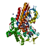
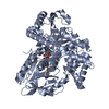
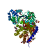
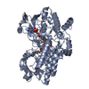
| Title | CRYSTAL STRUCTURE OF A NOVEL ADP-DEPENDENT GLUCOKINASE FROM THERMOCOCCUS LITORALIS | ||||||
 Components Components | ADP-DEPENDENT GLUCOKINASE | ||||||
 Keywords Keywords | TRANSFERASE / ALFA/BETA SANDWICHS / INDUCED-FITTING | ||||||
| Function / homology |  Function and homology information Function and homology informationADP-specific glucose/glucosamine kinase / ADP-specific glucokinase activity / Transferases; Transferring phosphorus-containing groups; Phosphotransferases with an alcohol group as acceptor / glucokinase activity / glycolytic process / glucose metabolic process / magnesium ion binding / cytoplasm Similarity search - Function | ||||||
| Biological species |   Thermococcus litoralis (archaea) Thermococcus litoralis (archaea) | ||||||
| Method |  X-RAY DIFFRACTION / SYNCHROTRON / Resolution: 2.3 Å X-RAY DIFFRACTION / SYNCHROTRON / Resolution: 2.3 Å | ||||||
 Authors Authors | Ito, S. / Fushinobu, S. / Yoshioka, I. / Koga, S. / Matsuzawa, H. / Wakagi, T. | ||||||
 Citation Citation | Journal: Structure / Year: 2001 Title: Structural Basis for the ADP-Specificity of a Novel Glucokinase from a Hyperthermophilic Archaeon Authors: Ito, S. / Fushinobu, S. / Yoshioka, I. / Koga, S. / Matsuzawa, H. / Wakagi, T. | ||||||
| History |
|
- Structure visualization
| Structure viewer | Molecule: MolmilJmol/JSmol |
|---|
- Downloads & links
-Download
| PDBx/mmCIF format | 1gc5.cif.gz | 106.2 KB | Display | PDBx/mmCIF format |
|---|---|---|---|---|
| PDB format | pdb1gc5.ent.gz | 82 KB | Display | PDB format |
| PDBx/mmJSON format | 1gc5.json.gz | Tree view | PDBx/mmJSON format | |
| Others |  Other downloads Other downloads |
-Validation report
| Arichive directory | https://data.pdbj.org/pub/pdb/validation_reports/gc/1gc5ftp://data.pdbj.org/pub/pdb/validation_reports/gc/1gc5 | HTTPS FTP |
|---|
-Related structure data
| Similar structure data |
|---|


-Links
 PDBj
PDBj- Assembly
| Deposited unit | 
| ||||||||
|---|---|---|---|---|---|---|---|---|---|
| 1 |
| ||||||||
| Unit cell |
| ||||||||
| Details | The biological assembly is monomer. |
-Components
| #1: Protein | Mass: 53618.086 Da / Num. of mol.: 1 Source method: isolated from a genetically manipulated source Source: (gene. exp.) Thermococcus litoralis (archaea) / Production host:  References: UniProt: Q7M537, ADP-specific glucose/glucosamine kinase |
|---|---|
| #2: Chemical | ChemComp-ADP /   Mass: 427.201 Da / Num. of mol.: 1 / Source method: obtained synthetically / Formula: C10H15N5O10P2 / Comment: ADP, energy-carrying molecule*YM Mass: 427.201 Da / Num. of mol.: 1 / Source method: obtained synthetically / Formula: C10H15N5O10P2 / Comment: ADP, energy-carrying molecule*YM |
| #3: Water | ChemComp-HOH /  Mass: 18.015 Da / Num. of mol.: 77 / Source method: isolated from a natural source / Formula: H2O Mass: 18.015 Da / Num. of mol.: 77 / Source method: isolated from a natural source / Formula: H2O |
-Experimental details
-Experiment
| Experiment | Method: X-RAY DIFFRACTION / Number of used crystals: 1 |
|---|
- Sample preparation
| Crystal | Density Matthews: 4.21 Å3/Da / Density % sol: 70.79 % | ||||||||||||||||||||||||||||||||||||
|---|---|---|---|---|---|---|---|---|---|---|---|---|---|---|---|---|---|---|---|---|---|---|---|---|---|---|---|---|---|---|---|---|---|---|---|---|---|
| Crystal grow | Temperature: 298 K / Method: vapor diffusion, hanging drop / pH: 4.8 Details: potassium chloride, citrate, pH 4.8, VAPOR DIFFUSION, HANGING DROP, temperature 298K | ||||||||||||||||||||||||||||||||||||
| Crystal grow | *PLUS Temperature: 25 ℃ | ||||||||||||||||||||||||||||||||||||
| Components of the solutions | *PLUS
|
-Data collection
| Diffraction | Mean temperature: 286 K |
|---|---|
| Diffraction source | Source: SYNCHROTRON / Site: Photon Factory  / Beamline: BL-6A / Wavelength: 1 / Beamline: BL-6A / Wavelength: 1 |
| Detector | Type: WEISSENBERG / Detector: DIFFRACTOMETER / Date: Jan 1, 1999 |
| Radiation | Protocol: SINGLE WAVELENGTH / Monochromatic (M) / Laue (L): M / Scattering type: x-ray |
| Radiation wavelength | Wavelength: 1 Å / Relative weight: 1 |
| Reflection | Resolution: 2.2→50 Å / Num. all: 167633 / Num. obs: 46477 / % possible obs: 77 % / Observed criterion σ(I): 1 / Redundancy: 3.3 % / Biso Wilson estimate: 33.3 Å2 / Rmerge(I) obs: 0.043 / Net I/σ(I): 18.6 |
| Reflection shell | Resolution: 2.2→2.28 Å / Rmerge(I) obs: 0.177 / % possible all: 45.8 |
| Reflection | *PLUS Lowest resolution: 50 Å / % possible obs: 77 % / Num. measured all: 167633 |
- Processing
| Software |
| ||||||||||||||||
|---|---|---|---|---|---|---|---|---|---|---|---|---|---|---|---|---|---|
| Refinement | Resolution: 2.3→6 Å / σ(F): 2 / Stereochemistry target values: Engh & Huber
| ||||||||||||||||
| Refinement step | Cycle: LAST / Resolution: 2.3→6 Å
| ||||||||||||||||
| Refine LS restraints |
| ||||||||||||||||
| Software | *PLUS Name: X-PLOR / Version: 3.851 / Classification: refinement | ||||||||||||||||
| Refinement | *PLUS Highest resolution: 2.3 Å / Lowest resolution: 6 Å / σ(F): 2 / Rfactor obs: 0.204 | ||||||||||||||||
| Solvent computation | *PLUS | ||||||||||||||||
| Displacement parameters | *PLUS | ||||||||||||||||
| Refine LS restraints | *PLUS
|
