 Movie
Movie Controller

[English] 日本語
 Yorodumi
- PDB-1gaj: CRYSTAL STRUCTURE OF A NUCLEOTIDE-FREE ATP-BINDING CASSETTE FROM ... -
+ Open data

- Basic information
| Entry | Database: PDB / ID: 1gaj | ||||||
|---|---|---|---|---|---|---|---|
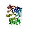
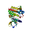
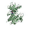
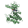
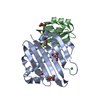
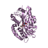
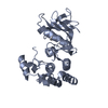
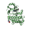
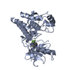
| Title | CRYSTAL STRUCTURE OF A NUCLEOTIDE-FREE ATP-BINDING CASSETTE FROM AN ABC TRANSPORTER | ||||||
 Components Components | HIGH-AFFINITY BRANCHED CHAIN AMINO ACID TRANSPORT ATP-BINDING PROTEIN | ||||||
 Keywords Keywords | TRANSPORT PROTEIN / ABC transporter / active transport / ATPase / nucleotide-binding domain | ||||||
| Function / homology |  Function and homology information Function and homology informationamino acid transport / ATP hydrolysis activity / ATP binding / plasma membrane Similarity search - Function | ||||||
| Biological species |   Methanocaldococcus jannaschii (archaea) Methanocaldococcus jannaschii (archaea) | ||||||
| Method |  X-RAY DIFFRACTION / SYNCHROTRON / MOLECULAR REPLACEMENT / Resolution: 2.5 Å X-RAY DIFFRACTION / SYNCHROTRON / MOLECULAR REPLACEMENT / Resolution: 2.5 Å | ||||||
 Authors Authors | Karpowich, N. / Yuan, Y.-R. / Dai, P.L. / Martsinkevich, O. / Millen, L. / Thomas, P.J. / Hunt, J.F. | ||||||
 Citation Citation | Journal: Structure / Year: 2001 Title: Crystal structures of the MJ1267 ATP binding cassette reveal an induced-fit effect at the ATPase active site of an ABC transporter. Authors: Karpowich, N. / Martsinkevich, O. / Millen, L. / Yuan, Y.R. / Dai, P.L. / MacVey, K. / Thomas, P.J. / Hunt, J.F. | ||||||
| History |
|
- Structure visualization
| Structure viewer | Molecule: MolmilJmol/JSmol |
|---|
- Downloads & links
-Download
| PDBx/mmCIF format | 1gaj.cif.gz | 67.8 KB | Display | PDBx/mmCIF format |
|---|---|---|---|---|
| PDB format | pdb1gaj.ent.gz | 50.1 KB | Display | PDB format |
| PDBx/mmJSON format | 1gaj.json.gz | Tree view | PDBx/mmJSON format | |
| Others |  Other downloads Other downloads |
-Validation report
| Arichive directory | https://data.pdbj.org/pub/pdb/validation_reports/ga/1gajftp://data.pdbj.org/pub/pdb/validation_reports/ga/1gaj | HTTPS FTP |
|---|
-Related structure data


-Links
 PDBj
PDBj

- Assembly
| Deposited unit | 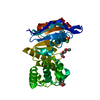
| ||||||||||||
|---|---|---|---|---|---|---|---|---|---|---|---|---|---|
| 1 |
| ||||||||||||
| 2 | x 24
| ||||||||||||
| Unit cell |
| ||||||||||||
| Components on special symmetry positions |
| ||||||||||||
| Details | The biological assembly is a monomer |
-Components
-Protein , 1 types, 1 molecules A
| #1: Protein | Mass: 29028.688 Da / Num. of mol.: 1 Source method: isolated from a genetically manipulated source Source: (gene. exp.) Methanocaldococcus jannaschii (archaea)Plasmid: PET28B / Production host:  |
|---|
-Non-polymers , 5 types, 141 molecules 


| #2: Chemical | ChemComp-SO4 /  Mass: 96.063 Da / Num. of mol.: 1 / Source method: obtained synthetically / Formula: SO4 Mass: 96.063 Da / Num. of mol.: 1 / Source method: obtained synthetically / Formula: SO4 | ||||||
|---|---|---|---|---|---|---|---|
| #3: Chemical |  Mass: 35.453 Da / Num. of mol.: 2 / Source method: obtained synthetically / Formula: Cl Mass: 35.453 Da / Num. of mol.: 2 / Source method: obtained synthetically / Formula: Cl#4: Chemical |  Mass: 74.122 Da / Num. of mol.: 3 / Source method: obtained synthetically / Formula: C4H10O Mass: 74.122 Da / Num. of mol.: 3 / Source method: obtained synthetically / Formula: C4H10O#5: Chemical |  Mass: 106.120 Da / Num. of mol.: 2 / Source method: obtained synthetically / Formula: C4H10O3 Mass: 106.120 Da / Num. of mol.: 2 / Source method: obtained synthetically / Formula: C4H10O3#6: Water | ChemComp-HOH / | Mass: 18.015 Da / Num. of mol.: 133 / Source method: isolated from a natural source / Formula: H2O |
-Experimental details
-Experiment
| Experiment | Method: X-RAY DIFFRACTION / Number of used crystals: 1 |
|---|
- Sample preparation
| Crystal | Density Matthews: 3.73 Å3/Da / Density % sol: 67.04 % | |||||||||||||||||||||||||||||||||||
|---|---|---|---|---|---|---|---|---|---|---|---|---|---|---|---|---|---|---|---|---|---|---|---|---|---|---|---|---|---|---|---|---|---|---|---|---|
| Crystal grow | Temperature: 294 K / Method: vapor diffusion, hanging drop / pH: 7.5 Details: HEPES-Cl, (NH4)2SO4, PEG 400, T-butanol, pH 7.5, VAPOR DIFFUSION, HANGING DROP, temperature 294K | |||||||||||||||||||||||||||||||||||
| Crystal grow | *PLUS Temperature: 21 ℃ | |||||||||||||||||||||||||||||||||||
| Components of the solutions | *PLUS
|
-Data collection
| Diffraction | Mean temperature: 130 K |
|---|---|
| Diffraction source | Source: SYNCHROTRON / Site: NSLS  / Beamline: X12B / Wavelength: 1.06884 Å / Beamline: X12B / Wavelength: 1.06884 Å |
| Detector | Type: ADSC QUANTUM 4 / Detector: CCD / Date: Jun 10, 2000 |
| Radiation | Monochromator: Si(111) / Protocol: SINGLE WAVELENGTH / Monochromatic (M) / Laue (L): M / Scattering type: x-ray |
| Radiation wavelength | Wavelength: 1.06884 Å / Relative weight: 1 |
| Reflection | Resolution: 2.5→40 Å / Num. all: 15956 / Num. obs: 13129 / % possible obs: 82.3 % / Observed criterion σ(F): 1 / Observed criterion σ(I): 3 / Redundancy: 39.9 % / Biso Wilson estimate: 54.4 Å2 / Rmerge(I) obs: 0.09 / Net I/σ(I): 44.16 |
| Reflection shell | Resolution: 2.5→2.61 Å / Redundancy: 19 % / Rmerge(I) obs: 0.43 / % possible all: 69 |
| Reflection | *PLUS Rmerge(I) obs: 0.09 |
- Processing
| Software |
| |||||||||||||||||||||||||
|---|---|---|---|---|---|---|---|---|---|---|---|---|---|---|---|---|---|---|---|---|---|---|---|---|---|---|
| Refinement | Method to determine structure: MOLECULAR REPLACEMENT / Resolution: 2.5→100 Å / σ(F): 1 / σ(I): 1 / Stereochemistry target values: X-PLOR 3.851
| |||||||||||||||||||||||||
| Refinement step | Cycle: LAST / Resolution: 2.5→100 Å
| |||||||||||||||||||||||||
| Software | *PLUS Name: X-PLOR / Version: 3.851 / Classification: refinement | |||||||||||||||||||||||||
| Refinement | *PLUS | |||||||||||||||||||||||||
| Solvent computation | *PLUS | |||||||||||||||||||||||||
| Displacement parameters | *PLUS | |||||||||||||||||||||||||
| Refine LS restraints | *PLUS
|
