 Movie
Movie Controller


+ Open data

- Basic information
| Entry | Database: PDB / ID: 1g5q | ||||||
|---|---|---|---|---|---|---|---|
| Title | EPID H67N COMPLEXED WITH SUBSTRATE PEPTIDE DSYTC | ||||||
 Components Components |
| ||||||
 Keywords Keywords | OXIDOREDUCTASE / alpha / beta protein / Rossman like fold | ||||||
| Function / homology |  Function and homology information Function and homology informationphosphopantothenoylcysteine decarboxylase activity / phosphopantothenoylcysteine decarboxylase complex / Lyases; Carbon-carbon lyases; Carboxy-lyases / coenzyme A biosynthetic process / FMN binding / killing of cells of another organism / defense response to bacterium / signaling receptor binding / extracellular region Similarity search - Function | ||||||
| Biological species |   Staphylococcus epidermidis (bacteria) Staphylococcus epidermidis (bacteria) | ||||||
| Method |  X-RAY DIFFRACTION / SYNCHROTRON / MOLECULAR REPLACEMENT / Resolution: 2.57 Å X-RAY DIFFRACTION / SYNCHROTRON / MOLECULAR REPLACEMENT / Resolution: 2.57 Å | ||||||
 Authors Authors | Blaesse, M. / Kupke, T. / Huber, R. / Steinbacher, S. | ||||||
 Citation Citation | Journal: EMBO J. / Year: 2000 Title: Crystal structure of the peptidyl-cysteine decarboxylase EpiD complexed with a pentapeptide substrate. Authors: Blaesse, M. / Kupke, T. / Huber, R. / Steinbacher, S. #1: Journal: J.Biol.Chem. / Year: 2000Title: Molecular characterization of lantibiotic synthesizing enzyme EpiD reveals a function for bacterial Dfp proteins in coenzyme A biosynthesis Authors: Kupke, T. / Uebele, M. / Schmid, D. / Jung, G. / Blaesse, M. / Steinbacher, S. | ||||||
| History |
|
- Structure visualization
| Structure viewer | Molecule: MolmilJmol/JSmol |
|---|
- Downloads & links
-Download
| PDBx/mmCIF format | 1g5q.cif.gz | 158.5 KB | Display | PDBx/mmCIF format |
|---|---|---|---|---|
| PDB format | pdb1g5q.ent.gz | 127.6 KB | Display | PDB format |
| PDBx/mmJSON format | 1g5q.json.gz | Tree view | PDBx/mmJSON format | |
| Others |  Other downloads Other downloads |
-Validation report
| Arichive directory | https://data.pdbj.org/pub/pdb/validation_reports/g5/1g5qftp://data.pdbj.org/pub/pdb/validation_reports/g5/1g5q | HTTPS FTP |
|---|
-Related structure data

-Links
 PDBj
PDBj- Assembly
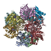
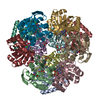
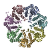
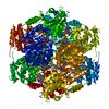
| Deposited unit | 
| |||||||||
|---|---|---|---|---|---|---|---|---|---|---|
| 1 | 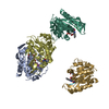
| |||||||||
| Unit cell |
| |||||||||
| Components on special symmetry positions |
| |||||||||
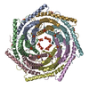
| Details | The biological assembly is a dodecamer generated from the tetramer in the asymmetric unit by the operations: -z+1, x-1/2, -y+1/2 and y+1/2, -z+1/2, -x+1. |
-Components
| #1: Protein | Mass: 20820.986 Da / Num. of mol.: 4 / Mutation: H67N Source method: isolated from a genetically manipulated source Source: (gene. exp.) Staphylococcus epidermidis (bacteria) / Strain: TUE3298 / Gene: EPID / Plasmid: PQE12 / Production host: #2: Protein/peptide | Mass: 587.601 Da / Num. of mol.: 4 / Fragment: C-TERMINUS / Mutation: N401D C404T / Source method: obtained synthetically / Details: The pentapeptide was chemically sythesized. / References: UniProt: P08136 #3: Chemical | ChemComp-FMN /   Mass: 456.344 Da / Num. of mol.: 4 / Source method: obtained synthetically / Formula: C17H21N4O9P Mass: 456.344 Da / Num. of mol.: 4 / Source method: obtained synthetically / Formula: C17H21N4O9P#4: Chemical |   Mass: 122.143 Da / Num. of mol.: 2 / Source method: obtained synthetically / Formula: C4H12NO3 / Comment: pH buffer*YM Mass: 122.143 Da / Num. of mol.: 2 / Source method: obtained synthetically / Formula: C4H12NO3 / Comment: pH buffer*YM#5: Water | ChemComp-HOH / |  Mass: 18.015 Da / Num. of mol.: 136 / Source method: isolated from a natural source / Formula: H2O Mass: 18.015 Da / Num. of mol.: 136 / Source method: isolated from a natural source / Formula: H2O |
|---|
-Experimental details
-Experiment
| Experiment | Method: X-RAY DIFFRACTION / Number of used crystals: 1 |
|---|
- Sample preparation
| Crystal | Density Matthews: 5.82 Å3/Da / Density % sol: 79 % | ||||||||||||||||||||||||
|---|---|---|---|---|---|---|---|---|---|---|---|---|---|---|---|---|---|---|---|---|---|---|---|---|---|
| Crystal grow | Temperature: 291 K / Method: vapor diffusion, sitting drop / pH: 6.5 Details: 100 mM MES/NaOH, 30 % MPD, 10 mM Peptide DSYTC, 3 mM DTT, 120 mM Glycine, pH 6.5, VAPOR DIFFUSION, SITTING DROP, temperature 291K | ||||||||||||||||||||||||
| Crystal grow | *PLUS Temperature: 18 ℃ / pH: 8 / Method: vapor diffusion | ||||||||||||||||||||||||
| Components of the solutions | *PLUS
|
-Data collection
| Diffraction | Mean temperature: 90 K |
|---|---|
| Diffraction source | Source: SYNCHROTRON / Site: MPG/DESY, HAMBURG  / Beamline: BW6 / Wavelength: 1.0499 Å / Beamline: BW6 / Wavelength: 1.0499 Å |
| Detector | Type: MARRESEARCH / Detector: CCD / Date: Mar 12, 2000 |
| Radiation | Monochromator: double crystal monochromator, Si(111) / Protocol: SINGLE WAVELENGTH / Monochromatic (M) / Laue (L): M / Scattering type: x-ray |
| Radiation wavelength | Wavelength: 1.0499 Å / Relative weight: 1 |
| Reflection | Resolution: 2.57→20 Å / Num. all: 151692 / Num. obs: 151692 / % possible obs: 93.2 % / Redundancy: 2.7 % / Biso Wilson estimate: 47.1 Å2 / Rmerge(I) obs: 0.068 / Rsym value: 6.8 / Net I/σ(I): 12.2 |
| Reflection shell | Resolution: 2.57→2.68 Å / Redundancy: 2.1 % / Rmerge(I) obs: 0.349 / Mean I/σ(I) obs: 2 / Num. unique all: 5205 / % possible all: 59.4 |
| Reflection | *PLUS Num. obs: 56352 / Num. measured all: 151692 |
| Reflection shell | *PLUS % possible obs: 59.4 % |
- Processing
| Software |
| ||||||||||||||||||||||||||||||||||||||||||||
|---|---|---|---|---|---|---|---|---|---|---|---|---|---|---|---|---|---|---|---|---|---|---|---|---|---|---|---|---|---|---|---|---|---|---|---|---|---|---|---|---|---|---|---|---|---|
| Refinement | Method to determine structure: MOLECULAR REPLACEMENT Starting model: tetramer from EpiD Resolution: 2.57→19.92 Å / Rfactor Rfree error: 0.004 / Data cutoff high absF: 3587123.43 / Data cutoff low absF: 0 / Isotropic thermal model: RESTRAINED / Cross valid method: THROUGHOUT / σ(F): 0 / Stereochemistry target values: Engh & Huber / Details: used maximum likelihood procedure
| ||||||||||||||||||||||||||||||||||||||||||||
| Displacement parameters | Biso mean: 50 Å2
| ||||||||||||||||||||||||||||||||||||||||||||
| Refine analyze |
| ||||||||||||||||||||||||||||||||||||||||||||
| Refinement step | Cycle: LAST / Resolution: 2.57→19.92 Å
| ||||||||||||||||||||||||||||||||||||||||||||
| Refine LS restraints |
| ||||||||||||||||||||||||||||||||||||||||||||
| Refine LS restraints NCS | NCS model details: CONSTRAINED | ||||||||||||||||||||||||||||||||||||||||||||
| LS refinement shell | Resolution: 2.57→2.66 Å / Rfactor Rfree error: 0.024 / Total num. of bins used: 10
| ||||||||||||||||||||||||||||||||||||||||||||
| Xplor file |
| ||||||||||||||||||||||||||||||||||||||||||||
| Software | *PLUS Name: CNS / Version: 1 / Classification: refinement | ||||||||||||||||||||||||||||||||||||||||||||
| Refinement | *PLUS σ(F): 0 / % reflection Rfree: 5.1 % / Rfactor obs: 0.209 | ||||||||||||||||||||||||||||||||||||||||||||
| Solvent computation | *PLUS | ||||||||||||||||||||||||||||||||||||||||||||
| Displacement parameters | *PLUS Biso mean: 50 Å2 | ||||||||||||||||||||||||||||||||||||||||||||
| Refine LS restraints | *PLUS
| ||||||||||||||||||||||||||||||||||||||||||||
| LS refinement shell | *PLUS Rfactor Rfree: 0.328 / % reflection Rfree: 4.8 % / Rfactor Rwork: 0.311 |
