 Movie
Movie Controller


+ Open data

- Basic information
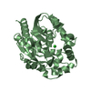
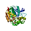
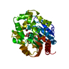
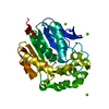
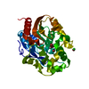
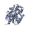
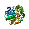
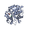
| Entry | Database: PDB / ID: 1g4h | ||||||
|---|---|---|---|---|---|---|---|
| Title | LINB COMPLEXED WITH BUTAN-1-OL | ||||||
 Components Components | 1,3,4,6-TETRACHLORO-1,4-CYCLOHEXADIENE HYDROLASE | ||||||
 Keywords Keywords | HYDROLASE / LinB Haloalkane dehalogenase halocarbon | ||||||
| Function / homology |  Function and homology information Function and homology informationhaloalkane dehalogenase / haloalkane dehalogenase activity / response to toxic substance / periplasmic space Similarity search - Function | ||||||
| Biological species |  Sphingomonas paucimobilis (bacteria) Sphingomonas paucimobilis (bacteria) | ||||||
| Method |  X-RAY DIFFRACTION / MOLECULAR REPLACEMENT / Resolution: 1.8 Å X-RAY DIFFRACTION / MOLECULAR REPLACEMENT / Resolution: 1.8 Å | ||||||
 Authors Authors | Oakley, A.J. / Prokop, Z. / Bohac, M. / Kmunicek, J. / Jedlicka, T. / Monincova, M. / Kuta-Smatanova, I. / Nagata, Y. / Damborsky, J. / Wilce, M.C.J. | ||||||
 Citation Citation | Journal: Biochemistry / Year: 2002 Title: Exploring the structure and activity of haloalkane dehalogenase from Sphingomonas paucimobilis UT26: evidence for product- and water-mediated inhibition. Authors: Oakley, A.J. / Prokop, Z. / Bohac, M. / Kmunicek, J. / Jedlicka, T. / Monincova, M. / Kuta-Smatanova, I. / Nagata, Y. / Damborsky, J. / Wilce, M.C. #1: Journal: Biochemistry / Year: 2000Title: Crystal structure of the haloalkane dehalogenase from Sphingomonas paucimobilis UT26 Authors: Marek, J. / Vevodova, J. / Smatanova, I.K. / Nagata, Y. / Svensson, L.A. / Newman, J. / Takagi, M. / Damborsky, J. | ||||||
| History |
|
- Structure visualization
| Structure viewer | Molecule: MolmilJmol/JSmol |
|---|
- Downloads & links
-Download
| PDBx/mmCIF format | 1g4h.cif.gz | 78 KB | Display | PDBx/mmCIF format |
|---|---|---|---|---|
| PDB format | pdb1g4h.ent.gz | 56.5 KB | Display | PDB format |
| PDBx/mmJSON format | 1g4h.json.gz | Tree view | PDBx/mmJSON format | |
| Others |  Other downloads Other downloads |
-Validation report
| Arichive directory | https://data.pdbj.org/pub/pdb/validation_reports/g4/1g4hftp://data.pdbj.org/pub/pdb/validation_reports/g4/1g4h | HTTPS FTP |
|---|
-Related structure data
| Related structure data | 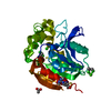 1g42C 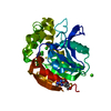 1g5fC 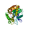 1cv2S C: citing same article ( S: Starting model for refinement |
|---|---|
| Similar structure data |


-Links
 PDBj
PDBj


- Assembly
| Deposited unit | 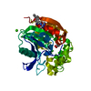
| ||||||||
|---|---|---|---|---|---|---|---|---|---|
| 1 |
| ||||||||
| Unit cell |
| ||||||||
| Components on special symmetry positions |
|
-Components
| #1: Protein | Mass: 33171.652 Da / Num. of mol.: 1 / Mutation: R291(2MR) Source method: isolated from a genetically manipulated source Source: (gene. exp.) Sphingomonas paucimobilis (bacteria) / Gene: LINB / Plasmid: PMLBH6 / Production host: References: UniProt: P51698, UniProt: D4Z2G1*PLUS, Hydrolases; Acting on halide bonds; In carbon-halide compounds | ||||||||
|---|---|---|---|---|---|---|---|---|---|
| #2: Chemical |   Mass: 40.078 Da / Num. of mol.: 2 / Source method: obtained synthetically / Formula: Ca Mass: 40.078 Da / Num. of mol.: 2 / Source method: obtained synthetically / Formula: Ca#3: Chemical | ChemComp-CL / |   Mass: 35.453 Da / Num. of mol.: 1 / Source method: obtained synthetically / Formula: Cl Mass: 35.453 Da / Num. of mol.: 1 / Source method: obtained synthetically / Formula: Cl#4: Chemical | 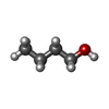  Mass: 74.122 Da / Num. of mol.: 3 / Source method: obtained synthetically / Formula: C4H10O Mass: 74.122 Da / Num. of mol.: 3 / Source method: obtained synthetically / Formula: C4H10O#5: Water | ChemComp-HOH / |  Mass: 18.015 Da / Num. of mol.: 225 / Source method: isolated from a natural source / Formula: H2O Mass: 18.015 Da / Num. of mol.: 225 / Source method: isolated from a natural source / Formula: H2OHas protein modification | Y | |
-Experimental details
-Experiment
| Experiment | Method: X-RAY DIFFRACTION / Number of used crystals: 1 |
|---|
- Sample preparation
| Crystal | Density Matthews: 2.03 Å3/Da / Density % sol: 26 % | ||||||||||||||||||||
|---|---|---|---|---|---|---|---|---|---|---|---|---|---|---|---|---|---|---|---|---|---|
| Crystal grow | Temperature: 298 K / Method: vapor diffusion, hanging drop / pH: 8.9 Details: 18-20% PEG 6000, 0.2 M Calcium Acetate, 0.1 M Tris-HCl pH 8.9, VAPOR DIFFUSION, HANGING DROP, temperature 298K | ||||||||||||||||||||
| Crystal grow | *PLUS | ||||||||||||||||||||
| Components of the solutions | *PLUS
|
-Data collection
| Diffraction | Mean temperature: 100 K |
|---|---|
| Diffraction source | Source: ROTATING ANODE / Type: RIGAKU RU200 / Wavelength: 1.5418 |
| Detector | Type: MARRESEARCH / Detector: AREA DETECTOR / Date: May 19, 2000 / Details: Ni Mirrors |
| Radiation | Monochromator: Ni Mirrors / Protocol: SINGLE WAVELENGTH / Monochromatic (M) / Laue (L): M / Scattering type: x-ray |
| Radiation wavelength | Wavelength: 1.5418 Å / Relative weight: 1 |
| Reflection | Resolution: 1.8→20 Å / Num. all: 24212 / Num. obs: 24212 / % possible obs: 94.3 % / Observed criterion σ(F): -3 / Observed criterion σ(I): -3 / Redundancy: 2.25 % / Biso Wilson estimate: 34.3 Å2 / Rmerge(I) obs: 0.04 / Net I/σ(I): 19.56 |
| Reflection shell | Resolution: 1.8→1.86 Å / Redundancy: 2.26 % / Rmerge(I) obs: 0.345 / Mean I/σ(I) obs: 3.5 / % possible all: 95.9 |
| Reflection | *PLUS Num. measured all: 57835 / Rmerge(I) obs: 0.04 |
| Reflection shell | *PLUS % possible obs: 95.9 % |
- Processing
| Software |
| |||||||||||||||||||||||||
|---|---|---|---|---|---|---|---|---|---|---|---|---|---|---|---|---|---|---|---|---|---|---|---|---|---|---|
| Refinement | Method to determine structure: MOLECULAR REPLACEMENT Starting model: PDB ENTRY 1CV2 Resolution: 1.8→17.45 Å / Rfactor Rfree error: 0.006 / Data cutoff high absF: 1082032.52 / Data cutoff low absF: 0 / Isotropic thermal model: RESTRAINED / Cross valid method: THROUGHOUT / σ(F): 0 / σ(I): 0 / Stereochemistry target values: Engh & Huber
| |||||||||||||||||||||||||
| Solvent computation | Solvent model: FLAT MODEL / Bsol: 78.08 Å2 / ksol: 0.429 e/Å3 | |||||||||||||||||||||||||
| Displacement parameters | Biso mean: 34.6 Å2
| |||||||||||||||||||||||||
| Refine analyze |
| |||||||||||||||||||||||||
| Refinement step | Cycle: LAST / Resolution: 1.8→17.45 Å
| |||||||||||||||||||||||||
| Refine LS restraints |
| |||||||||||||||||||||||||
| LS refinement shell | Resolution: 1.8→1.91 Å / Rfactor Rfree error: 0.019 / Total num. of bins used: 6
| |||||||||||||||||||||||||
| Xplor file |
| |||||||||||||||||||||||||
| Software | *PLUS Name: CNS / Version: 1 / Classification: refinement | |||||||||||||||||||||||||
| Refinement | *PLUS σ(F): 0 / % reflection Rfree: 5.1 % | |||||||||||||||||||||||||
| Solvent computation | *PLUS | |||||||||||||||||||||||||
| Displacement parameters | *PLUS Biso mean: 34.6 Å2 | |||||||||||||||||||||||||
| Refine LS restraints | *PLUS
| |||||||||||||||||||||||||
| LS refinement shell | *PLUS Rfactor Rfree: 0.273 / % reflection Rfree: 5.1 % / Rfactor Rwork: 0.269 |
