 Movie
Movie Controller


+ Open data

- Basic information
| Entry | Database: PDB / ID: 1g1b | ||||||
|---|---|---|---|---|---|---|---|
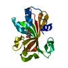
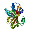
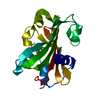
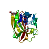
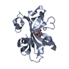
| Title | CHORISMATE LYASE (WILD-TYPE) WITH BOUND PRODUCT | ||||||
 Components Components | CHORISMATE LYASE | ||||||
 Keywords Keywords | LYASE / 6-stranded-antiparallel-sheet topology=(123654) | ||||||
| Function / homology |  Function and homology information Function and homology informationchorismate lyase / chorismate lyase activity / pyruvate biosynthetic process / ubiquinone biosynthetic process / cytosol Similarity search - Function | ||||||
| Biological species |  | ||||||
| Method |  X-RAY DIFFRACTION / Resolution: 1.99 Å X-RAY DIFFRACTION / Resolution: 1.99 Å | ||||||
 Authors Authors | Gallagher, D.T. / Mayhew, M. / Holden, M.J. / Kim, K.J. / Howard, A. / Vilker, V.L. | ||||||
 Citation Citation | Journal: Proteins / Year: 2001 Title: The crystal structure of chorismate lyase shows a new fold and a tightly retained product. Authors: Gallagher, D.T. / Mayhew, M. / Holden, M.J. / Howard, A. / Kim, K.J. / Vilker, V.L. #1: Journal: J.Struct.Biol. / Year: 2000Title: Crystallization and 1.1-A Diffraction of Chorismate Lyase from Escherichia coli Authors: Stover, C. / Mayhew, M.P. / Holden, M.J. / Howard, A. / Gallagher, D.T. | ||||||
| History |
|
- Structure visualization
| Structure viewer | Molecule: MolmilJmol/JSmol |
|---|
- Downloads & links
-Download
| PDBx/mmCIF format | 1g1b.cif.gz | 82.6 KB | Display | PDBx/mmCIF format |
|---|---|---|---|---|
| PDB format | pdb1g1b.ent.gz | 62.1 KB | Display | PDB format |
| PDBx/mmJSON format | 1g1b.json.gz | Tree view | PDBx/mmJSON format | |
| Others |  Other downloads Other downloads |
-Validation report
| Arichive directory | https://data.pdbj.org/pub/pdb/validation_reports/g1/1g1bftp://data.pdbj.org/pub/pdb/validation_reports/g1/1g1b | HTTPS FTP |
|---|
-Related structure data


-Links
 PDBj
PDBj- Assembly
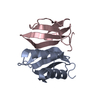
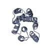
| Deposited unit | 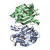
| ||||||||
|---|---|---|---|---|---|---|---|---|---|
| 1 | 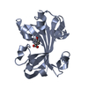
| ||||||||
| 2 | 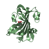
| ||||||||
| Unit cell |
| ||||||||
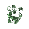
| Details | Asymmetric unit contains two monomers, each with bound product. Biological unit is a monomer. |
-Components
| #1: Protein | Mass: 18667.686 Da / Num. of mol.: 2 / Source method: isolated from a natural source / Source: (natural) #2: Chemical |   Mass: 138.121 Da / Num. of mol.: 2 / Source method: obtained synthetically / Formula: C7H6O3 Mass: 138.121 Da / Num. of mol.: 2 / Source method: obtained synthetically / Formula: C7H6O3#3: Water | ChemComp-HOH / |  Mass: 18.015 Da / Num. of mol.: 250 / Source method: isolated from a natural source / Formula: H2O Mass: 18.015 Da / Num. of mol.: 250 / Source method: isolated from a natural source / Formula: H2O |
|---|
-Experimental details
-Experiment
| Experiment | Method: X-RAY DIFFRACTION / Number of used crystals: 1 |
|---|
- Sample preparation
| Crystal | Density Matthews: 2.11 Å3/Da / Density % sol: 41.68 % | |||||||||||||||||||||||||
|---|---|---|---|---|---|---|---|---|---|---|---|---|---|---|---|---|---|---|---|---|---|---|---|---|---|---|
| Crystal grow | Temperature: 298 K / Method: vapor diffusion, hanging drop / pH: 7 Details: 100 mM Hepes buffer, 22% (v/v) PEG 4K, 5% (v/v) isopropanol, pH 7.0, VAPOR DIFFUSION, HANGING DROP, temperature 298.0K | |||||||||||||||||||||||||
| Crystal grow | *PLUS Temperature: 4 ℃ / Method: vapor diffusion / Details: Stover, C., (2000) J. Struct. Biol., 129, 96. | |||||||||||||||||||||||||
| Components of the solutions | *PLUS
|
-Data collection
| Diffraction | Mean temperature: 100 K |
|---|---|
| Diffraction source | Source: ROTATING ANODE / Type: SIEMENS / Wavelength: 1.5418 |
| Detector | Type: MARRESEARCH / Detector: IMAGE PLATE / Date: Mar 20, 1998 |
| Radiation | Protocol: SINGLE WAVELENGTH / Monochromatic (M) / Laue (L): M / Scattering type: x-ray |
| Radiation wavelength | Wavelength: 1.5418 Å / Relative weight: 1 |
| Reflection | Resolution: 2→20 Å / Num. all: 20385 / Num. obs: 20385 / % possible obs: 93.9 % / Observed criterion σ(F): 0 / Observed criterion σ(I): 0 / Redundancy: 5.5 % / Biso Wilson estimate: 15.4 Å2 / Rmerge(I) obs: 0.062 / Net I/σ(I): 18.1 |
| Reflection shell | Resolution: 1.98→2.07 Å / Redundancy: 1.5 % / Rmerge(I) obs: 0.22 / Num. unique all: 2093 / % possible all: 77.7 |
| Reflection | *PLUS % possible obs: 94 % / Num. measured all: 111649 / Rmerge(I) obs: 0.06 |
| Reflection shell | *PLUS Highest resolution: 2 Å / % possible obs: 78 % |
- Processing
| Software |
| |||||||||||||||||||||||||
|---|---|---|---|---|---|---|---|---|---|---|---|---|---|---|---|---|---|---|---|---|---|---|---|---|---|---|
| Refinement | Resolution: 1.99→7.99 Å / Rfactor Rfree error: 0.008 / Data cutoff high absF: 423436.69 / Data cutoff low absF: 0 / Isotropic thermal model: RESTRAINED / Cross valid method: THROUGHOUT / σ(F): 0 / σ(I): 0 / Stereochemistry target values: Engh & Huber
| |||||||||||||||||||||||||
| Solvent computation | Solvent model: FLAT MODEL / Bsol: 113.1 Å2 / ksol: 0.62 e/Å3 | |||||||||||||||||||||||||
| Displacement parameters | Biso mean: 16.3 Å2
| |||||||||||||||||||||||||
| Refine analyze |
| |||||||||||||||||||||||||
| Refinement step | Cycle: LAST / Resolution: 1.99→7.99 Å
| |||||||||||||||||||||||||
| Refine LS restraints |
| |||||||||||||||||||||||||
| LS refinement shell | Resolution: 1→1.05 Å / Total num. of bins used: 8 /
| |||||||||||||||||||||||||
| Xplor file |
| |||||||||||||||||||||||||
| Software | *PLUS Name: CNS / Version: 1 / Classification: refinement | |||||||||||||||||||||||||
| Refinement | *PLUS Highest resolution: 2 Å / Lowest resolution: 8 Å / Num. reflection Rfree: 1018 / % reflection Rfree: 5 % / Rfactor Rfree: 0.27 / Rfactor Rwork: 0.2 | |||||||||||||||||||||||||
| Solvent computation | *PLUS | |||||||||||||||||||||||||
| Displacement parameters | *PLUS | |||||||||||||||||||||||||
| Refine LS restraints | *PLUS
| |||||||||||||||||||||||||
| LS refinement shell | *PLUS Highest resolution: 2 Å / Lowest resolution: 2.07 Å / Rfactor Rfree: 0.28 / Rfactor Rwork: 0.19 / Num. reflection obs: 1817 |
