 Movie
Movie Controller

[English] 日本語
 Yorodumi
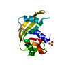
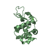
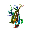
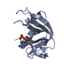
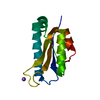
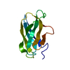
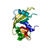
- PDB-1fxd: REFINED CRYSTAL STRUCTURE OF FERREDOXIN II FROM DESULFOVIBRIO GIG... -
+ Open data

- Basic information
| Entry | Database: PDB / ID: 1fxd | |||||||||
|---|---|---|---|---|---|---|---|---|---|---|
| Title | REFINED CRYSTAL STRUCTURE OF FERREDOXIN II FROM DESULFOVIBRIO GIGAS AT 1.7 ANGSTROMS | |||||||||
 Components Components | FERREDOXIN II | |||||||||
 Keywords Keywords | ELECTRON TRANSFER(IRON-SULFUR) | |||||||||
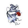
| Function / homology |  Function and homology information Function and homology information3 iron, 4 sulfur cluster binding / electron transfer activity / iron ion binding Similarity search - Function | |||||||||
| Biological species |  Desulfovibrio gigas (bacteria) Desulfovibrio gigas (bacteria) | |||||||||
| Method |  X-RAY DIFFRACTION / Resolution: 1.7 Å X-RAY DIFFRACTION / Resolution: 1.7 Å | |||||||||
 Authors Authors | Kissinger, C.R. / Sieker, L.C. / Adman, E.T. / Jensen, L.H. | |||||||||
 Citation Citation | Journal: J.Mol.Biol. / Year: 1991 Title: Refined crystal structure of ferredoxin II from Desulfovibrio gigas at 1.7 A. Authors: Kissinger, C.R. / Sieker, L.C. / Adman, E.T. / Jensen, L.H. #1: Journal: FEBS Lett. / Year: 1989Title: The Crystal Structure of the Three-Iron Ferredoxin II from Desulfovibrio Gigas Authors: Kissinger, C.R. / Adman, E.T. / Sieker, L.C. / Jensen, L.H. / Legall, J. #2: Journal: J.Am.Chem.Soc. / Year: 1988Title: Structure of the 3Fe-4S Cluster in Desulfovibrio Gigas Ferredoxin II Authors: Kissinger, C.R. / Adman, E.T. / Sieker, L.C. / Jensen, L.H. #3: Journal: J.Mol.Biol. / Year: 1984Title: Crystallization and Preliminary X-Ray Diffraction Study of the 3-Fe Ferredoxin II from the Bacterium Desulfovibrio Gigas Authors: Sieker, L.C. / Adman, E.T. / Jensen, L.H. / Legall, J. | |||||||||
| History |
|
- Structure visualization
| Structure viewer | Molecule: MolmilJmol/JSmol |
|---|
- Downloads & links
-Download
| PDBx/mmCIF format | 1fxd.cif.gz | 24 KB | Display | PDBx/mmCIF format |
|---|---|---|---|---|
| PDB format | pdb1fxd.ent.gz | 14.8 KB | Display | PDB format |
| PDBx/mmJSON format | 1fxd.json.gz | Tree view | PDBx/mmJSON format | |
| Others |  Other downloads Other downloads |
-Validation report
| Arichive directory | https://data.pdbj.org/pub/pdb/validation_reports/fx/1fxdftp://data.pdbj.org/pub/pdb/validation_reports/fx/1fxd | HTTPS FTP |
|---|
-Related structure data
| Similar structure data |
|---|

-Links
 PDBj
PDBj


- Assembly
| Deposited unit | 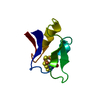
| ||||||||||||||||||
|---|---|---|---|---|---|---|---|---|---|---|---|---|---|---|---|---|---|---|---|
| 1 | 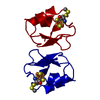
| ||||||||||||||||||
| Unit cell |
| ||||||||||||||||||
| Components on special symmetry positions |
|
-Components
| #1: Protein | Mass: 6310.010 Da / Num. of mol.: 1 Source method: isolated from a genetically manipulated source Source: (gene. exp.) Desulfovibrio gigas (bacteria) / References: UniProt: P00209 |
|---|---|
| #2: Chemical | ChemComp-F3S /   Mass: 295.795 Da / Num. of mol.: 1 / Source method: obtained synthetically / Formula: Fe3S4 Mass: 295.795 Da / Num. of mol.: 1 / Source method: obtained synthetically / Formula: Fe3S4 |
| #3: Water | ChemComp-HOH /  Mass: 18.015 Da / Num. of mol.: 56 / Source method: isolated from a natural source / Formula: H2O Mass: 18.015 Da / Num. of mol.: 56 / Source method: isolated from a natural source / Formula: H2O |
| Has protein modification | Y |
-Experimental details
-Experiment
| Experiment | Method: X-RAY DIFFRACTION |
|---|
- Sample preparation
| Crystal | Density Matthews: 1.88 Å3/Da / Density % sol: 34.44 % |
|---|---|
| Crystal grow | *PLUS Method: other / Details: Sieker, L.C., (1984) J. Mol. Biol., 179, 151. |
-Data collection
| Radiation | Scattering type: x-ray |
|---|---|
| Radiation wavelength | Relative weight: 1 |
| Reflection | *PLUS Highest resolution: 1.7 Å / Lowest resolution: 2.3 Å / Num. obs: 6228 / Rmerge(I) obs: 0.09 |
- Processing
| Software | Name: PROFFT / Classification: refinement | |||||||||||||||||||||||||||||||||||||||||||||||||||||||||||||||
|---|---|---|---|---|---|---|---|---|---|---|---|---|---|---|---|---|---|---|---|---|---|---|---|---|---|---|---|---|---|---|---|---|---|---|---|---|---|---|---|---|---|---|---|---|---|---|---|---|---|---|---|---|---|---|---|---|---|---|---|---|---|---|---|---|
| Refinement | Rfactor obs: 0.157 / Highest resolution: 1.7 Å | |||||||||||||||||||||||||||||||||||||||||||||||||||||||||||||||
| Refinement step | Cycle: LAST / Highest resolution: 1.7 Å
| |||||||||||||||||||||||||||||||||||||||||||||||||||||||||||||||
| Refine LS restraints |
| |||||||||||||||||||||||||||||||||||||||||||||||||||||||||||||||
| Refinement | *PLUS Lowest resolution: 8 Å / Num. reflection obs: 4508 | |||||||||||||||||||||||||||||||||||||||||||||||||||||||||||||||
| Solvent computation | *PLUS | |||||||||||||||||||||||||||||||||||||||||||||||||||||||||||||||
| Displacement parameters | *PLUS | |||||||||||||||||||||||||||||||||||||||||||||||||||||||||||||||
| Refine LS restraints | *PLUS
|
