 Movie
Movie Controller

[English] 日本語
 Yorodumi
- PDB-1frf: CRYSTAL STRUCTURE OF THE NI-FE HYDROGENASE FROM DESULFOVIBRIO FRU... -
+ Open data

- Basic information
| Entry | Database: PDB / ID: 1frf | ||||||||||||
|---|---|---|---|---|---|---|---|---|---|---|---|---|---|
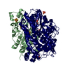
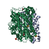
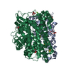
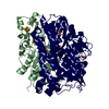
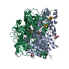
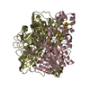
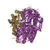
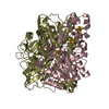
| Title | CRYSTAL STRUCTURE OF THE NI-FE HYDROGENASE FROM DESULFOVIBRIO FRUCTOSOVORANS | ||||||||||||
 Components Components | ([NI-FE] HYDROGENASE) x 2 | ||||||||||||
 Keywords Keywords | OXIDOREDUCTASE / NI-FE HYDROGENASE | ||||||||||||
| Function / homology |  Function and homology information Function and homology informationcytochrome-c3 hydrogenase / cytochrome-c3 hydrogenase activity / ferredoxin hydrogenase complex / [Ni-Fe] hydrogenase complex / ferredoxin hydrogenase activity / anaerobic respiration / 3 iron, 4 sulfur cluster binding / nickel cation binding / 4 iron, 4 sulfur cluster binding / periplasmic space ...cytochrome-c3 hydrogenase / cytochrome-c3 hydrogenase activity / ferredoxin hydrogenase complex / [Ni-Fe] hydrogenase complex / ferredoxin hydrogenase activity / anaerobic respiration / 3 iron, 4 sulfur cluster binding / nickel cation binding / 4 iron, 4 sulfur cluster binding / periplasmic space / electron transfer activity / membrane / metal ion binding Similarity search - Function | ||||||||||||
| Biological species |  Desulfovibrio fructosovorans (bacteria) Desulfovibrio fructosovorans (bacteria) | ||||||||||||
| Method |  X-RAY DIFFRACTION / SYNCHROTRON / MOLECULAR REPLACEMENT / Resolution: 2.7 Å X-RAY DIFFRACTION / SYNCHROTRON / MOLECULAR REPLACEMENT / Resolution: 2.7 Å | ||||||||||||
 Authors Authors | Montet, Y. / Volbeda, A. / Piras, C. / Hatchikian, E.C. / Frey, M. / Fontecilla, J.C. | ||||||||||||
 Citation Citation | Journal: Proc.Natl.Acad.Sci.USA / Year: 1998 Title: 3Fe-4S] to [4Fe-4S] cluster conversion in Desulfovibrio fructosovorans [NiFe] hydrogenase by site-directed mutagenesis Authors: Rousset, M. / Montet, Y. / Guigliarelli, B. / Forget, N. / Asso, M. / Bertrand, P. / Fontecilla-Camps, J.C. / Hatchikian, E.C. #1: Journal: Nat.Struct.Biol. / Year: 1997Title: Gas Access to the Active Site of Ni-Fe Hydrogeases Probed by X-Ray Crystallography and Molecular Dynamics Authors: Montet, Y. / Amara, P. / Volbeda, A. / Vernede, X. / Hatchikian, E.C. / Field, M.J. / Frey, M. / Fontecilla-Camps, J.C. | ||||||||||||
| History |
|
- Structure visualization
| Structure viewer | Molecule: MolmilJmol/JSmol |
|---|
- Downloads & links
-Download
| PDBx/mmCIF format | 1frf.cif.gz | 170.5 KB | Display | PDBx/mmCIF format |
|---|---|---|---|---|
| PDB format | pdb1frf.ent.gz | 132.8 KB | Display | PDB format |
| PDBx/mmJSON format | 1frf.json.gz | Tree view | PDBx/mmJSON format | |
| Others |  Other downloads Other downloads |
-Validation report
| Arichive directory | https://data.pdbj.org/pub/pdb/validation_reports/fr/1frfftp://data.pdbj.org/pub/pdb/validation_reports/fr/1frf | HTTPS FTP |
|---|
-Related structure data
| Related structure data |  1frvS S: Starting model for refinement |
|---|---|
| Similar structure data |

-Links
 PDBj
PDBj


- Assembly
| Deposited unit | 
| ||||||||
|---|---|---|---|---|---|---|---|---|---|
| 1 |
| ||||||||
| Unit cell |
| ||||||||
| Noncrystallographic symmetry (NCS) | NCS oper: (Code: given Matrix: (0.394555, 0.745792, 0.536769), Vector: |
-Components
-Protein , 2 types, 2 molecules SL
| #1: Protein | [ Mass: 28422.400 Da / Num. of mol.: 1 / Fragment: SMALL CHAIN / Source method: isolated from a natural source / Details: DSM 3604 / Source: (natural) Desulfovibrio fructosovorans (bacteria) / Cellular location: PERIPLASM / Strain: WILD TYPE / References: UniProt: P18187, 1.18.99.1 |
|---|---|
| #2: Protein | [ Mass: 61669.488 Da / Num. of mol.: 1 / Fragment: LARGE CHAIN / Source method: isolated from a natural source / Details: DSM 3604 / Source: (natural) Desulfovibrio fructosovorans (bacteria) / Cellular location: PERIPLASM / Strain: WILD TYPE / References: UniProt: P18188, 1.18.99.1 |
-Non-polymers , 6 types, 138 molecules 


| #3: Chemical |  Mass: 351.640 Da / Num. of mol.: 2 / Source method: obtained synthetically / Formula: Fe4S4 Mass: 351.640 Da / Num. of mol.: 2 / Source method: obtained synthetically / Formula: Fe4S4#4: Chemical | ChemComp-F3S / |  Mass: 295.795 Da / Num. of mol.: 1 / Source method: obtained synthetically / Formula: Fe3S4 Mass: 295.795 Da / Num. of mol.: 1 / Source method: obtained synthetically / Formula: Fe3S4#5: Chemical | ChemComp-FE / |  Mass: 55.845 Da / Num. of mol.: 1 / Source method: obtained synthetically / Formula: Fe Mass: 55.845 Da / Num. of mol.: 1 / Source method: obtained synthetically / Formula: Fe#6: Chemical | ChemComp-NI / |  Mass: 58.693 Da / Num. of mol.: 1 / Source method: obtained synthetically / Formula: Ni Mass: 58.693 Da / Num. of mol.: 1 / Source method: obtained synthetically / Formula: Ni#7: Chemical | ChemComp-MG / |  Mass: 24.305 Da / Num. of mol.: 1 / Source method: obtained synthetically / Formula: Mg Mass: 24.305 Da / Num. of mol.: 1 / Source method: obtained synthetically / Formula: Mg#8: Water | ChemComp-HOH / | Mass: 18.015 Da / Num. of mol.: 132 / Source method: isolated from a natural source / Formula: H2O |
|---|
-Details
| Has protein modification | Y |
|---|
-Experimental details
-Experiment
| Experiment | Method: X-RAY DIFFRACTION / Number of used crystals: 1 |
|---|
- Sample preparation
| Crystal | Density Matthews: 2.2 Å3/Da / Density % sol: 44.1 % | |||||||||||||||||||||||||
|---|---|---|---|---|---|---|---|---|---|---|---|---|---|---|---|---|---|---|---|---|---|---|---|---|---|---|
| Crystal grow | pH: 6.4 Details: PEG 6000 28%, MES 100MM PH 6,4 25MM OCTYL-BETA-D-GLUCOPYRANOSIDE, pH 6.4 | |||||||||||||||||||||||||
| Crystal grow | *PLUS Method: unknown | |||||||||||||||||||||||||
| Components of the solutions | *PLUS
|
-Data collection
| Diffraction | Mean temperature: 110 K |
|---|---|
| Diffraction source | Source: SYNCHROTRON / Site: ESRF  / Beamline: BM02 / Wavelength: 0.9794 / Beamline: BM02 / Wavelength: 0.9794 |
| Detector | Date: Oct 1, 1996 / Details: MIRRORS |
| Radiation | Protocol: SINGLE WAVELENGTH / Monochromatic (M) / Laue (L): M / Scattering type: x-ray |
| Radiation wavelength | Wavelength: 0.9794 Å / Relative weight: 1 |
| Reflection | Resolution: 2.7→38.7 Å / Num. obs: 43607 / % possible obs: 66.7 % / Observed criterion σ(I): 2 / Redundancy: 2.8 % / Rsym value: 0.061 / Net I/σ(I): 10.2 |
| Reflection shell | Resolution: 2.7→2.77 Å / Redundancy: 1.3 % / Mean I/σ(I) obs: 2 / Rsym value: 0.32 / % possible all: 56.4 |
| Reflection | *PLUS Num. obs: 33794 / % possible obs: 74 % / Num. measured all: 79013 / Rmerge(I) obs: 0.077 |
- Processing
| Software |
| ||||||||||||||||||||||||||||||||||||||||||||||||||||||||||||
|---|---|---|---|---|---|---|---|---|---|---|---|---|---|---|---|---|---|---|---|---|---|---|---|---|---|---|---|---|---|---|---|---|---|---|---|---|---|---|---|---|---|---|---|---|---|---|---|---|---|---|---|---|---|---|---|---|---|---|---|---|---|
| Refinement | Method to determine structure: MOLECULAR REPLACEMENT Starting model: 1FRV Resolution: 2.7→12 Å / Data cutoff high absF: 0 / Data cutoff low absF: 0.001 / σ(F): 2 Details: THERE ARE THREE MOLECULES PER ASYMETRIC UNIT. IN ALL REFINEMENT STEPS, THE STRICT NON CRYSTALLOGRAPHIC SYMMETRY WAS USED. THERE IS NO ELECTRON DENSITY FOR RESIDUES 1 - 5 OF THE LARGE SUBUNIT ...Details: THERE ARE THREE MOLECULES PER ASYMETRIC UNIT. IN ALL REFINEMENT STEPS, THE STRICT NON CRYSTALLOGRAPHIC SYMMETRY WAS USED. THERE IS NO ELECTRON DENSITY FOR RESIDUES 1 - 5 OF THE LARGE SUBUNIT AND RESIDUES 1 - 3 FOR THE SMALL ONE. SOME ATOMS HAVE OCCUPANCIES LESS THAN 1.0 : THEY BELONG TO LATERAL CHAINS WHICH ARE DISORDERED , AS THEY ARE LOCATED ON THE SURFACE OF THE PROTEIN.
| ||||||||||||||||||||||||||||||||||||||||||||||||||||||||||||
| Displacement parameters | Biso mean: 12.48 Å2 | ||||||||||||||||||||||||||||||||||||||||||||||||||||||||||||
| Refinement step | Cycle: LAST / Resolution: 2.7→12 Å
| ||||||||||||||||||||||||||||||||||||||||||||||||||||||||||||
| Refine LS restraints |
| ||||||||||||||||||||||||||||||||||||||||||||||||||||||||||||
| Refine LS restraints NCS | NCS model details: STRICT | ||||||||||||||||||||||||||||||||||||||||||||||||||||||||||||
| Xplor file | Serial no: 1 / Param file: PARHCSDX.PRO / Topol file: TOPHCSDX.PRO | ||||||||||||||||||||||||||||||||||||||||||||||||||||||||||||
| Software | *PLUS Name: X-PLOR / Version: 3.1 / Classification: refinement | ||||||||||||||||||||||||||||||||||||||||||||||||||||||||||||
| Refinement | *PLUS Lowest resolution: 12 Å / σ(F): 2 / % reflection Rfree: 5 % / Rfactor Rwork: 0.22 | ||||||||||||||||||||||||||||||||||||||||||||||||||||||||||||
| Solvent computation | *PLUS | ||||||||||||||||||||||||||||||||||||||||||||||||||||||||||||
| Displacement parameters | *PLUS | ||||||||||||||||||||||||||||||||||||||||||||||||||||||||||||
| Refine LS restraints | *PLUS
|
