 Movie
Movie Controller


+ Open data

- Basic information
| Entry | Database: PDB / ID: 1fl1 | ||||||
|---|---|---|---|---|---|---|---|
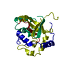
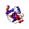
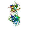
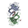
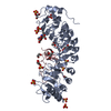
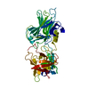
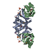
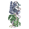
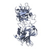
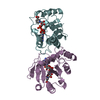
| Title | KSHV PROTEASE | ||||||
 Components Components | PROTEASE | ||||||
 Keywords Keywords | VIRAL PROTEIN / serine protease / antiviral drug design / capsid maturation / endopeptidase / assemblin | ||||||
| Function / homology |  Function and homology information Function and homology informationassemblin / nuclear capsid assembly / viral release from host cell / host cell cytoplasm / serine-type endopeptidase activity / host cell nucleus / proteolysis / identical protein binding Similarity search - Function | ||||||
| Biological species |   Human herpesvirus 8 Human herpesvirus 8 | ||||||
| Method |  X-RAY DIFFRACTION / SYNCHROTRON / Resolution: 2.2 Å X-RAY DIFFRACTION / SYNCHROTRON / Resolution: 2.2 Å | ||||||
 Authors Authors | Reiling, K.K. / Pray, T.R. / Craik, C.S. / Stroud, R.M. | ||||||
 Citation Citation | Journal: Biochemistry / Year: 2000 Title: Functional consequences of the Kaposi's sarcoma-associated herpesvirus protease structure: regulation of activity and dimerization by conserved structural elements. Authors: Reiling, K.K. / Pray, T.R. / Craik, C.S. / Stroud, R.M. #1: Journal: Acta Crystallogr.,Sect.D / Year: 1998Title: Crystallography & NMR System: A new software suite for macromolecular structure determination Authors: Brunger, A.T. / Adams, P.D. / Clore, G.M. / Delano, W.L. / Gros, P. / Grosse-Kunstleve, R. / Jiang, J.-S. / Kuszewski, J. / Nilges, M. / Pannu, N.S. / Read, R.J. / Rice, L.M. / Simonson, T. / Warren, G. | ||||||
| History |
|
- Structure visualization
| Structure viewer | Molecule: MolmilJmol/JSmol |
|---|
- Downloads & links
-Download
| PDBx/mmCIF format | 1fl1.cif.gz | 91.6 KB | Display | PDBx/mmCIF format |
|---|---|---|---|---|
| PDB format | pdb1fl1.ent.gz | 69.4 KB | Display | PDB format |
| PDBx/mmJSON format | 1fl1.json.gz | Tree view | PDBx/mmJSON format | |
| Others |  Other downloads Other downloads |
-Validation report
| Summary document | 1fl1_validation.pdf.gz | 409.4 KB | Display | wwPDB validaton report |
|---|---|---|---|---|
| Full document | 1fl1_full_validation.pdf.gz | 413.1 KB | Display | |
| Data in XML | 1fl1_validation.xml.gz | 10.1 KB | Display | |
| Data in CIF | 1fl1_validation.cif.gz | 15.4 KB | Display | |
| Arichive directory | https://data.pdbj.org/pub/pdb/validation_reports/fl/1fl1ftp://data.pdbj.org/pub/pdb/validation_reports/fl/1fl1 | HTTPS FTP |
-Related structure data
| Related structure data | |
|---|---|
| Similar structure data |


-Links
 PDBj
PDBj- Assembly
| Deposited unit | 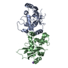
| ||||||||
|---|---|---|---|---|---|---|---|---|---|
| 1 |
| ||||||||
| 2 | 
| ||||||||
| Unit cell |
| ||||||||
| Details | The biological active enzyme is a homo-dimer constructed from chain A and B related by a Non-crystalographic two-fold. |
-Components
| #1: Protein | Mass: 25229.938 Da / Num. of mol.: 2 / Mutation: S204G Source method: isolated from a genetically manipulated source Source: (gene. exp.) Human herpesvirus 8 / Genus: Rhadinovirus / Plasmid: PQE30 / Production host:  #2: Chemical | ChemComp-K / |   Mass: 39.098 Da / Num. of mol.: 1 / Source method: obtained synthetically / Formula: K Mass: 39.098 Da / Num. of mol.: 1 / Source method: obtained synthetically / Formula: K#3: Water | ChemComp-HOH / |  Mass: 18.015 Da / Num. of mol.: 169 / Source method: isolated from a natural source / Formula: H2O Mass: 18.015 Da / Num. of mol.: 169 / Source method: isolated from a natural source / Formula: H2O |
|---|
-Experimental details
-Experiment
| Experiment | Method: X-RAY DIFFRACTION / Number of used crystals: 1 |
|---|
- Sample preparation
| Crystal | Density Matthews: 2.65 Å3/Da / Density % sol: 53.6 % | ||||||||||||||||||||||||||||||||||||||||||||||||||||||||||||||||||||||||||||||||||||||||||
|---|---|---|---|---|---|---|---|---|---|---|---|---|---|---|---|---|---|---|---|---|---|---|---|---|---|---|---|---|---|---|---|---|---|---|---|---|---|---|---|---|---|---|---|---|---|---|---|---|---|---|---|---|---|---|---|---|---|---|---|---|---|---|---|---|---|---|---|---|---|---|---|---|---|---|---|---|---|---|---|---|---|---|---|---|---|---|---|---|---|---|---|
| Crystal grow | Temperature: 298 K / Method: vapor diffusion, hanging drop / pH: 7.5 Details: 22% PEG-2K, 100 mM Tris-HCl, 10% glycerol, 190 mM LiSO4, pH 7.5, VAPOR DIFFUSION, HANGING DROP, temperature 25K | ||||||||||||||||||||||||||||||||||||||||||||||||||||||||||||||||||||||||||||||||||||||||||
| Crystal | *PLUS Density % sol: 53.7 % | ||||||||||||||||||||||||||||||||||||||||||||||||||||||||||||||||||||||||||||||||||||||||||
| Crystal grow | *PLUS pH: 7 | ||||||||||||||||||||||||||||||||||||||||||||||||||||||||||||||||||||||||||||||||||||||||||
| Components of the solutions | *PLUS
|
-Data collection
| Diffraction | Mean temperature: 100 K |
|---|---|
| Diffraction source | Source: SYNCHROTRON / Site: SSRL  / Beamline: BL9-1 / Wavelength: 0.98 / Beamline: BL9-1 / Wavelength: 0.98 |
| Detector | Type: MARRESEARCH / Detector: IMAGE PLATE / Date: Aug 3, 1999 |
| Radiation | Protocol: SINGLE WAVELENGTH / Monochromatic (M) / Laue (L): M / Scattering type: x-ray |
| Radiation wavelength | Wavelength: 0.98 Å / Relative weight: 1 |
| Reflection | Resolution: 2.2→28.282 Å / Num. all: 28220 / Num. obs: 28034 / % possible obs: 99.4 % / Observed criterion σ(F): -3 / Observed criterion σ(I): -3 / Redundancy: 5 % / Biso Wilson estimate: 41.658 Å2 / Rmerge(I) obs: 0.062 / Net I/σ(I): 17.2 |
| Reflection shell | Resolution: 2.2→2.22 Å / Redundancy: 5 % / Rmerge(I) obs: 0.351 / Num. unique all: 905 / % possible all: 99.5 |
- Processing
| Software |
| ||||||||||||||||||||
|---|---|---|---|---|---|---|---|---|---|---|---|---|---|---|---|---|---|---|---|---|---|
| Refinement | Resolution: 2.2→28 Å / σ(F): 0 / σ(I): 0 Stereochemistry target values: Cambridge Data Base model structures (R. A. Engh and R. Huber, Acta Cryst. Sect. A., 1991). Details: RESIDUES 182 AND 207 HAD DENSITY ONLY TO THE CG AND WERE MODELED AS SERINES DURING REFINEMENT.
| ||||||||||||||||||||
| Refinement step | Cycle: LAST / Resolution: 2.2→28 Å
| ||||||||||||||||||||
| Refine LS restraints |
| ||||||||||||||||||||
| Software | *PLUS Name: CNS / Version: 1 / Classification: refinement | ||||||||||||||||||||
| Refinement | *PLUS Lowest resolution: 28 Å / σ(F): 0 | ||||||||||||||||||||
| Solvent computation | *PLUS | ||||||||||||||||||||
| Displacement parameters | *PLUS | ||||||||||||||||||||
| Refine LS restraints | *PLUS
|
