 Movie
Movie Controller

[English] 日本語
 Yorodumi
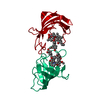
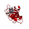
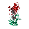
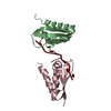
- PDB-1fkd: FK-506 BINDING PROTEIN: THREE-DIMENSIONAL STRUCTURE OF THE COMPLE... -
+ Open data

- Basic information
| Entry | Database: PDB / ID: 1fkd | ||||||
|---|---|---|---|---|---|---|---|
| Title | FK-506 BINDING PROTEIN: THREE-DIMENSIONAL STRUCTURE OF THE COMPLEX WITH THE ANTAGONIST L-685,818 | ||||||
 Components Components | FK506 BINDING PROTEIN | ||||||
 Keywords Keywords | CIS-TRANS ISOMERASE | ||||||
| Function / homology |  Function and homology information Function and homology informationmacrolide binding / activin receptor binding / regulation of skeletal muscle contraction by regulation of release of sequestered calcium ion / cytoplasmic side of membrane / transforming growth factor beta receptor binding / TGFBR1 LBD Mutants in Cancer / type I transforming growth factor beta receptor binding / negative regulation of activin receptor signaling pathway / signaling receptor inhibitor activity / heart trabecula formation ...macrolide binding / activin receptor binding / regulation of skeletal muscle contraction by regulation of release of sequestered calcium ion / cytoplasmic side of membrane / transforming growth factor beta receptor binding / TGFBR1 LBD Mutants in Cancer / type I transforming growth factor beta receptor binding / negative regulation of activin receptor signaling pathway / signaling receptor inhibitor activity / heart trabecula formation / I-SMAD binding / regulation of amyloid precursor protein catabolic process / terminal cisterna / ryanodine receptor complex / 'de novo' protein folding / ventricular cardiac muscle tissue morphogenesis / FK506 binding / TGF-beta receptor signaling activates SMADs / mTORC1-mediated signalling / Calcineurin activates NFAT / regulation of immune response / heart morphogenesis / supramolecular fiber organization / regulation of cardiac muscle contraction by regulation of the release of sequestered calcium ion / sarcoplasmic reticulum membrane / T cell activation / sarcoplasmic reticulum / peptidylprolyl isomerase / peptidyl-prolyl cis-trans isomerase activity / TGF-beta receptor signaling in EMT (epithelial to mesenchymal transition) / calcium channel regulator activity / negative regulation of transforming growth factor beta receptor signaling pathway / protein maturation / Z disc / SARS-CoV-1 activates/modulates innate immune responses / regulation of protein localization / protein refolding / protein folding / amyloid fibril formation / Potential therapeutics for SARS / transmembrane transporter binding / positive regulation of canonical NF-kappaB signal transduction / membrane / cytoplasm / cytosol Similarity search - Function | ||||||
| Biological species |  Homo sapiens (human) Homo sapiens (human) | ||||||
| Method |  X-RAY DIFFRACTION / Resolution: 1.72 Å X-RAY DIFFRACTION / Resolution: 1.72 Å | ||||||
 Authors Authors | Becker, J.W. / Rotonda, J. / Mckeever, B.M. | ||||||
 Citation Citation | Journal: J.Biol.Chem. / Year: 1993 Title: FK-506-binding protein: three-dimensional structure of the complex with the antagonist L-685,818. Authors: Becker, J.W. / Rotonda, J. / McKeever, B.M. / Chan, H.K. / Marcy, A.I. / Wiederrecht, G. / Hermes, J.D. / Springer, J.P. #1: Journal: J.Exp.Med. / Year: 1992Title: The Immunosuppressive and Toxic Effects of Fk-506 are Mechanistically Related: Pharmacology of a Novel Antagonist of Fk-506 and Rapamycin Authors: Dumont, F.J. / Staruch, M.J. / Koprak, S.L. / Siekierka, J.J. / Lin, C.S. / Harrison, R. / Sewell, T. / Kindt, V.M. / Beattie, T.R. / Wyvratt, M. / Sigal, N.H. | ||||||
| History |
| ||||||
| Remark 700 | SHEET THE SHEET STRUCTURE OF THIS MOLECULE IS BIFURCATED. IN ORDER TO REPRESENT THIS FEATURE IN THE ...SHEET THE SHEET STRUCTURE OF THIS MOLECULE IS BIFURCATED. IN ORDER TO REPRESENT THIS FEATURE IN THE SHEET RECORDS BELOW, TWO SHEETS ARE DEFINED. STRANDS 1, 2, 3, AND 4 OF A1 AND A2 BELOW ARE IDENTICAL WHILE STRAND 5 IS DIFFERENT. |
- Structure visualization
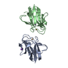
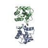
| Structure viewer | Molecule: MolmilJmol/JSmol |
|---|
- Downloads & links
-Download
| PDBx/mmCIF format | 1fkd.cif.gz | 36.3 KB | Display | PDBx/mmCIF format |
|---|---|---|---|---|
| PDB format | pdb1fkd.ent.gz | 24.4 KB | Display | PDB format |
| PDBx/mmJSON format | 1fkd.json.gz | Tree view | PDBx/mmJSON format | |
| Others |  Other downloads Other downloads |
-Validation report
| Arichive directory | https://data.pdbj.org/pub/pdb/validation_reports/fk/1fkdftp://data.pdbj.org/pub/pdb/validation_reports/fk/1fkd | HTTPS FTP |
|---|
-Related structure data


-Links
 PDBj
PDBj


- Assembly
| Deposited unit | 
| ||||||||
|---|---|---|---|---|---|---|---|---|---|
| 1 | 
| ||||||||
| 2 | 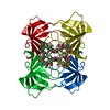
| ||||||||
| Unit cell |
| ||||||||
| Components on special symmetry positions |
|
-Components
| #1: Protein | Mass: 11836.508 Da / Num. of mol.: 1 Source method: isolated from a genetically manipulated source Source: (gene. exp.) Homo sapiens (human) / References: UniProt: P62942 |
|---|---|
| #2: Chemical | ChemComp-818 /   Mass: 808.007 Da / Num. of mol.: 1 / Source method: obtained synthetically / Formula: C43H69NO13 Mass: 808.007 Da / Num. of mol.: 1 / Source method: obtained synthetically / Formula: C43H69NO13 |
| #3: Water | ChemComp-HOH /  Mass: 18.015 Da / Num. of mol.: 64 / Source method: isolated from a natural source / Formula: H2O Mass: 18.015 Da / Num. of mol.: 64 / Source method: isolated from a natural source / Formula: H2O |
-Experimental details
-Experiment
| Experiment | Method: X-RAY DIFFRACTION |
|---|
- Sample preparation
| Crystal | Density Matthews: 1.99 Å3/Da / Density % sol: 38.05 % |
|---|---|
| Crystal grow | *PLUS pH: 4.5 / Method: vapor diffusion |
| Components of the solutions | *PLUS Conc.: 57 %sat / Chemical formula: (NH4)2SO4 |
-Data collection
| Radiation | Scattering type: x-ray |
|---|---|
| Radiation wavelength | Relative weight: 1 |
| Reflection | *PLUS Highest resolution: 1.72 Å / Num. obs: 11113 / Num. measured all: 64522 / Rmerge(I) obs: 0.0594 |
- Processing
| Software | Name: PROLSQ / Classification: refinement | ||||||||||||||||||||||||||||||||||||||||||||||||||||||||||||||||||||||||||||||||||||
|---|---|---|---|---|---|---|---|---|---|---|---|---|---|---|---|---|---|---|---|---|---|---|---|---|---|---|---|---|---|---|---|---|---|---|---|---|---|---|---|---|---|---|---|---|---|---|---|---|---|---|---|---|---|---|---|---|---|---|---|---|---|---|---|---|---|---|---|---|---|---|---|---|---|---|---|---|---|---|---|---|---|---|---|---|---|
| Refinement | Resolution: 1.72→8 Å / σ(F): 0 /
| ||||||||||||||||||||||||||||||||||||||||||||||||||||||||||||||||||||||||||||||||||||
| Refinement step | Cycle: LAST / Resolution: 1.72→8 Å
| ||||||||||||||||||||||||||||||||||||||||||||||||||||||||||||||||||||||||||||||||||||
| Refine LS restraints |
| ||||||||||||||||||||||||||||||||||||||||||||||||||||||||||||||||||||||||||||||||||||
| Refinement | *PLUS Highest resolution: 1.72 Å / Lowest resolution: 8 Å / Num. reflection all: 10312 / σ(F): 0 / Rfactor all: 0.181 | ||||||||||||||||||||||||||||||||||||||||||||||||||||||||||||||||||||||||||||||||||||
| Solvent computation | *PLUS | ||||||||||||||||||||||||||||||||||||||||||||||||||||||||||||||||||||||||||||||||||||
| Displacement parameters | *PLUS |
