 Movie
Movie Controller


+ Open data

- Basic information
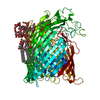
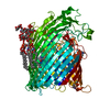
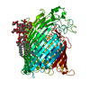
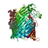
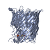
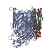
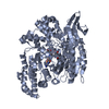
| Entry | Database: PDB / ID: 1fi1 | |||||||||
|---|---|---|---|---|---|---|---|---|---|---|
| Title | FhuA in complex with lipopolysaccharide and rifamycin CGP4832 | |||||||||
 Components Components | FERRICHROME-IRON RECEPTOR | |||||||||
 Keywords Keywords | METAL TRANSPORT / Outer membrane protein / TonB-dependent receptor / FhuA / siderophore receptor / integral membrane protein / lipopolysaccharide / rifamycin CGP 4832 / beta-barrel / antibiotic | |||||||||
| Function / homology |  Function and homology information Function and homology informationsiderophore-iron import into cell / siderophore transmembrane transport / siderophore uptake transmembrane transporter activity / transmembrane transporter complex / virion binding / toxic substance binding / cell outer membrane / signaling receptor activity / intracellular iron ion homeostasis / iron ion binding ...siderophore-iron import into cell / siderophore transmembrane transport / siderophore uptake transmembrane transporter activity / transmembrane transporter complex / virion binding / toxic substance binding / cell outer membrane / signaling receptor activity / intracellular iron ion homeostasis / iron ion binding / protein domain specific binding / membrane Similarity search - Function | |||||||||
| Biological species |  | |||||||||
| Method |  X-RAY DIFFRACTION / SYNCHROTRON / Resolution: 2.9 Å X-RAY DIFFRACTION / SYNCHROTRON / Resolution: 2.9 Å | |||||||||
 Authors Authors | Ferguson, A.D. / Koedding, J. / Boes, C. / Walker, G. / Coulton, J.W. / Diederichs, K. / Braun, V. / Welte, W. | |||||||||
 Citation Citation | Journal: Structure / Year: 2001 Title: Active transport of an antibiotic rifamycin derivative by the outer-membrane protein FhuA. Authors: Ferguson, A.D. / Kodding, J. / Walker, G. / Bos, C. / Coulton, J.W. / Diederichs, K. / Braun, V. / Welte, W. #1: Journal: Structure / Year: 2000Title: A conserved structural motif for lipopolysaccharide recognition by procaryotic and eucaryotic proteins Authors: Ferguson, A.D. / Welte, W. / Hofmann, E. / Lindner, B. / Holst, O. / Coulton, J.W. / Diederichs, K. #2: Journal: Protein Sci. / Year: 2000Title: Crystal structure of the antibiotic albomycin in complex with the outer membrane transporter FhuA Authors: Ferguson, A.D. / Braun, V. / Fiedler, H.-P. / Coulton, J.W. / Diederichs, K. / Welte, W. #3: Journal: Science / Year: 1998Title: Siderophore-mediated iron transport: Crystal structure of FhuA with bound lipopolysaccharide Authors: Ferguson, A.D. / Hofmann, E. / Coulton, J.W. / Diederichs, K. / Welte, W. | |||||||||
| History |
|
- Structure visualization
| Structure viewer | Molecule: MolmilJmol/JSmol |
|---|
- Downloads & links
-Download
| PDBx/mmCIF format | 1fi1.cif.gz | 171.3 KB | Display | PDBx/mmCIF format |
|---|---|---|---|---|
| PDB format | pdb1fi1.ent.gz | 130.2 KB | Display | PDB format |
| PDBx/mmJSON format | 1fi1.json.gz | Tree view | PDBx/mmJSON format | |
| Others |  Other downloads Other downloads |
-Validation report
| Arichive directory | https://data.pdbj.org/pub/pdb/validation_reports/fi/1fi1ftp://data.pdbj.org/pub/pdb/validation_reports/fi/1fi1 | HTTPS FTP |
|---|
-Related structure data
| Related structure data | |
|---|---|
| Similar structure data |


-Links
 PDBj
PDBj
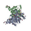
- Assembly
| Deposited unit | 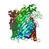
| ||||||||
|---|---|---|---|---|---|---|---|---|---|
| 1 |
| ||||||||
| Unit cell |
|
-Components
-Protein / Sugars , 2 types, 2 molecules A
| #1: Protein | Mass: 78230.078 Da / Num. of mol.: 1 Mutation: HEXAHISTIDINE TAG PLUS FIVE LINKER RESIDUES HAVE BEEN GENETICALLY INSERTED AFTER RESIDUE 405 OF THE MATURE SEQUENCE Source method: isolated from a genetically manipulated source Source: (gene. exp.) |
|---|---|
| #2: Polysaccharide | alpha-D-glucopyranose-(1-2)-alpha-D-glucopyranose-(1-3)-[alpha-D-galactopyranose-(1-6)]alpha-D- ...alpha-D-glucopyranose-(1-2)-alpha-D-glucopyranose-(1-3)-[alpha-D-galactopyranose-(1-6)]alpha-D-glucopyranose-(1-3)-[L-glycero-alpha-D-manno-heptopyranose-(1-7)]L-glycero-alpha-D-manno-heptopyranose-(1-3)-L-glycero-alpha-D-manno-heptopyranose-(1-5)-[3-deoxy-alpha-D-manno-oct-2-ulopyranosonic acid-(2-4)]3-deoxy-alpha-D-manno-oct-2-ulopyranosonic acid-(2-6)-2-amino-2,3-dideoxy-alpha-D-glucoyranose-(1-6)-2-amino-2-deoxy-alpha-D-glucopyranose Source method: isolated from a genetically manipulated source |
-Non-polymers , 9 types, 194 molecules 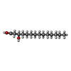


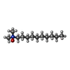

| #3: Chemical | ChemComp-FTT /  Mass: 244.370 Da / Num. of mol.: 6 Mass: 244.370 Da / Num. of mol.: 6Source method: isolated from a genetically manipulated source Formula: C14H28O3 #4: Chemical |  Mass: 94.971 Da / Num. of mol.: 2 / Source method: obtained synthetically / Formula: PO4 Mass: 94.971 Da / Num. of mol.: 2 / Source method: obtained synthetically / Formula: PO4#5: Chemical | ChemComp-NI / |  Mass: 58.693 Da / Num. of mol.: 1 / Source method: obtained synthetically / Formula: Ni Mass: 58.693 Da / Num. of mol.: 1 / Source method: obtained synthetically / Formula: Ni#6: Chemical | ChemComp-NA / |  Mass: 22.990 Da / Num. of mol.: 1 / Source method: obtained synthetically / Formula: Na Mass: 22.990 Da / Num. of mol.: 1 / Source method: obtained synthetically / Formula: Na#7: Chemical | ChemComp-MG / |  Mass: 24.305 Da / Num. of mol.: 1 / Source method: obtained synthetically / Formula: Mg Mass: 24.305 Da / Num. of mol.: 1 / Source method: obtained synthetically / Formula: Mg#8: Chemical |  Mass: 173.943 Da / Num. of mol.: 2 / Source method: obtained synthetically / Formula: O7P2 Mass: 173.943 Da / Num. of mol.: 2 / Source method: obtained synthetically / Formula: O7P2#9: Chemical | ChemComp-RIF / |  Mass: 936.051 Da / Num. of mol.: 1 / Source method: obtained synthetically / Formula: C49H65N3O15 Mass: 936.051 Da / Num. of mol.: 1 / Source method: obtained synthetically / Formula: C49H65N3O15#10: Chemical | ChemComp-DDQ / |  Mass: 201.349 Da / Num. of mol.: 1 / Source method: obtained synthetically / Formula: C12H27NO Mass: 201.349 Da / Num. of mol.: 1 / Source method: obtained synthetically / Formula: C12H27NO#11: Water | ChemComp-HOH / | Mass: 18.015 Da / Num. of mol.: 179 / Source method: isolated from a natural source / Formula: H2O |
|---|
-Details
| Has protein modification | Y |
|---|
-Experimental details
-Experiment
| Experiment | Method: X-RAY DIFFRACTION / Number of used crystals: 1 |
|---|
- Sample preparation
| Crystal | Density Matthews: 4.84 Å3/Da / Density % sol: 74.6 % | ||||||||||||||||||||
|---|---|---|---|---|---|---|---|---|---|---|---|---|---|---|---|---|---|---|---|---|---|
| Crystal grow | Temperature: 291 K / Method: vapor diffusion, hanging drop / pH: 6.4 Details: 100 mM sodium cacodylate, 12% PEG 2,000 MME, 3% PEG 200, 1% cis-inositol, 20% glycerol, 1 mM rifamycin CGP 4832, pH 6.4, VAPOR DIFFUSION, HANGING DROP, temperature 291K | ||||||||||||||||||||
| Crystal grow | *PLUS Temperature: 18 ℃ / Details: Ferguson, A.D., (1998) Protein Sci., 7, 1636. | ||||||||||||||||||||
| Components of the solutions | *PLUS
|
-Data collection
| Diffraction | Mean temperature: 100 K |
|---|---|
| Diffraction source | Source: SYNCHROTRON / Site: ELETTRA  / Beamline: 5.2R / Wavelength: 1 / Beamline: 5.2R / Wavelength: 1 |
| Detector | Type: MARRESEARCH / Detector: AREA DETECTOR / Date: Jul 3, 1999 |
| Radiation | Protocol: SINGLE WAVELENGTH / Monochromatic (M) / Laue (L): M / Scattering type: x-ray |
| Radiation wavelength | Wavelength: 1 Å / Relative weight: 1 |
| Reflection | Resolution: 2.9→30 Å / Num. obs: 33363 / % possible obs: 99.9 % / Observed criterion σ(F): 1 / Observed criterion σ(I): 1 / Redundancy: 4.83 % / Biso Wilson estimate: 23.2 Å2 / Rmerge(I) obs: 0.072 / Net I/σ(I): 17.2 |
| Reflection shell | Resolution: 2.9→3.08 Å / Redundancy: 4.83 % / Rmerge(I) obs: 0.246 / Num. unique all: 33363 / % possible all: 99.8 |
| Reflection | *PLUS Num. measured all: 161168 |
| Reflection shell | *PLUS % possible obs: 99.8 % / Mean I/σ(I) obs: 4.2 |
- Processing
| Software |
| ||||||||||||||||||||||||||||||||||||||||
|---|---|---|---|---|---|---|---|---|---|---|---|---|---|---|---|---|---|---|---|---|---|---|---|---|---|---|---|---|---|---|---|---|---|---|---|---|---|---|---|---|---|
| Refinement | Resolution: 2.9→30 Å / Rfactor Rfree error: 0.007 / Data cutoff high absF: 467052.59 / Data cutoff low absF: 0 / Isotropic thermal model: RESTRAINED / Cross valid method: THROUGHOUT / σ(F): 0 / σ(I): 0 / Stereochemistry target values: Engh & Huber
| ||||||||||||||||||||||||||||||||||||||||
| Solvent computation | Solvent model: FLAT MODEL / Bsol: 60.07 Å2 / ksol: 0.3 e/Å3 | ||||||||||||||||||||||||||||||||||||||||
| Displacement parameters | Biso mean: 68.4 Å2
| ||||||||||||||||||||||||||||||||||||||||
| Refine analyze |
| ||||||||||||||||||||||||||||||||||||||||
| Refinement step | Cycle: LAST / Resolution: 2.9→30 Å
| ||||||||||||||||||||||||||||||||||||||||
| Refine LS restraints |
| ||||||||||||||||||||||||||||||||||||||||
| LS refinement shell | Resolution: 2.9→3.08 Å / Rfactor Rfree error: 0.027 / Total num. of bins used: 6
| ||||||||||||||||||||||||||||||||||||||||
| Xplor file |
| ||||||||||||||||||||||||||||||||||||||||
| Software | *PLUS Name: CNS / Classification: refinement | ||||||||||||||||||||||||||||||||||||||||
| Refinement | *PLUS Lowest resolution: 30 Å / σ(F): 0 / % reflection Rfree: 4.7 % | ||||||||||||||||||||||||||||||||||||||||
| Solvent computation | *PLUS | ||||||||||||||||||||||||||||||||||||||||
| Displacement parameters | *PLUS Biso mean: 68.4 Å2 | ||||||||||||||||||||||||||||||||||||||||
| Refine LS restraints | *PLUS
| ||||||||||||||||||||||||||||||||||||||||
| LS refinement shell | *PLUS Rfactor Rfree: 0.387 / % reflection Rfree: 3.6 % / Rfactor Rwork: 0.329 |
