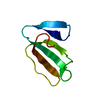
 Movie
Movie Controller


+ Open data

- Basic information
| Entry | Database: PDB / ID: 1ffj | ||||||
|---|---|---|---|---|---|---|---|
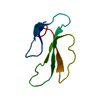
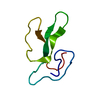
| Title | NMR STRUCTURE OF CARDIOTOXIN IN DPC-MICELLE | ||||||
 Components Components | CYTOTOXIN 2 | ||||||
 Keywords Keywords | TOXIN / all-beta sheet protein / membrane perturbation | ||||||
| Function / homology |  Function and homology information Function and homology informationother organism cell membrane / toxin activity / killing of cells of another organism / extracellular region / membrane Similarity search - Function | ||||||
| Biological species |  Naja oxiana (Central Asian cobra) Naja oxiana (Central Asian cobra) | ||||||
| Method | SOLUTION NMR / torsion angle dynamics | ||||||
 Authors Authors | Dubovskii, P.V. / Dementieva, D.V. / Bocharov, E.V. / Utkin, Y.N. / Arseniev, A.S. | ||||||
 Citation Citation | Journal: J.Mol.Biol. / Year: 2001 Title: Membrane binding motif of the P-type cardiotoxin. Authors: Dubovskii, P.V. / Dementieva, D.V. / Bocharov, E.V. / Utkin, Y.N. / Arseniev, A.S. #1: Journal: Eur.J.Biochem. / Year: 1999Title: Two forms of Cytotoxin II (cardiotoxin) from Naja naja oxiana in Aqueous Solution. Spatial Structures with Tightly Bound Water Molecules Authors: Dementieva, D.V. / Bocharov, E.V. / Arseniev, A.S. | ||||||
| History |
|
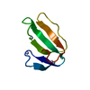
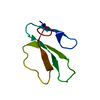
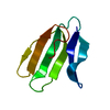
- Structure visualization
| Structure viewer | Molecule: MolmilJmol/JSmol |
|---|
- Downloads & links
-Download
| PDBx/mmCIF format | 1ffj.cif.gz | 353.3 KB | Display | PDBx/mmCIF format |
|---|---|---|---|---|
| PDB format | pdb1ffj.ent.gz | 294.8 KB | Display | PDB format |
| PDBx/mmJSON format | 1ffj.json.gz | Tree view | PDBx/mmJSON format | |
| Others |  Other downloads Other downloads |
-Validation report
| Arichive directory | https://data.pdbj.org/pub/pdb/validation_reports/ff/1ffjftp://data.pdbj.org/pub/pdb/validation_reports/ff/1ffj | HTTPS FTP |
|---|
-Related structure data
| Related structure data | |
|---|---|
| Similar structure data |


-Links
 PDBj
PDBj

- Assembly
| Deposited unit | 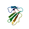
| |||||||||
|---|---|---|---|---|---|---|---|---|---|---|
| 1 |
| |||||||||
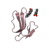
| NMR ensembles |
|
-Components
| #1: Protein | Mass: 6648.238 Da / Num. of mol.: 1 / Fragment: 1 / Source method: isolated from a natural source / Source: (natural) Naja oxiana (Central Asian cobra) / Secretion: VENOM / References: UniProt: P01441 |
|---|---|
| #2: Water | ChemComp-HOH /  Mass: 18.015 Da / Num. of mol.: 2 / Source method: isolated from a natural source / Formula: H2O Mass: 18.015 Da / Num. of mol.: 2 / Source method: isolated from a natural source / Formula: H2O |
| Has protein modification | Y |
-Experimental details
-Experiment
| Experiment | Method: SOLUTION NMR | ||||||||||||||||||||||||||||||||||||||||||||
|---|---|---|---|---|---|---|---|---|---|---|---|---|---|---|---|---|---|---|---|---|---|---|---|---|---|---|---|---|---|---|---|---|---|---|---|---|---|---|---|---|---|---|---|---|---|
| NMR experiment |
| ||||||||||||||||||||||||||||||||||||||||||||
| NMR details | Text: This structure was determined using standard 2D homonuclear techniques. Sites of tightly bound water molecules were determined as in Dementieva et al., Eur.J.Biochem.1999,263,152-162. |
- Sample preparation
| Details |
| |||||||||||||||
|---|---|---|---|---|---|---|---|---|---|---|---|---|---|---|---|---|
| Sample conditions |
| |||||||||||||||
| Crystal grow | *PLUS Method: other / Details: NMR |
-NMR measurement
| NMR spectrometer | Type: Varian UNITY / Manufacturer: Varian / Model: UNITY / Field strength: 600 MHz |
|---|
- Processing
| NMR software |
| ||||||||||||||||||||
|---|---|---|---|---|---|---|---|---|---|---|---|---|---|---|---|---|---|---|---|---|---|
| Refinement | Method: torsion angle dynamics / Software ordinal: 1 Details: structures are based on a total of 368 NOE-derived constraints, 154 dihedral angle restraints, 248 distance restraints from hydrogen bonds and disulfides | ||||||||||||||||||||
| NMR representative | Selection criteria: fewest violations | ||||||||||||||||||||
| NMR ensemble | Conformer selection criteria: target function / Conformers calculated total number: 220 / Conformers submitted total number: 20 |
