 Movie
Movie Controller


+ Open data

- Basic information
| Entry | Database: PDB / ID: 1ff9 | ||||||
|---|---|---|---|---|---|---|---|
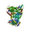
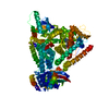
| Title | APO SACCHAROPINE REDUCTASE | ||||||
 Components Components | SACCHAROPINE REDUCTASE | ||||||
 Keywords Keywords | OXIDOREDUCTASE / LYSINE BIOSYNTHESIS / ALPHA-AMINOADIPATE PATHWAY / SACCHAROPINE REDUCTASE / DEHYDROGENASE | ||||||
| Function / homology |  Function and homology information Function and homology informationsaccharopine dehydrogenase (NADP+, L-glutamate-forming) / saccharopine dehydrogenase (NADP+, L-glutamate-forming) activity / lysine biosynthetic process via aminoadipic acid / cytoplasm Similarity search - Function | ||||||
| Biological species |  Magnaporthe grisea (fungus) Magnaporthe grisea (fungus) | ||||||
| Method |  X-RAY DIFFRACTION / SYNCHROTRON / Resolution: 2 Å X-RAY DIFFRACTION / SYNCHROTRON / Resolution: 2 Å | ||||||
 Authors Authors | Johansson, E. / Steffens, J.J. / Lindqvist, Y. / Schneider, G. | ||||||
 Citation Citation | Journal: Structure Fold.Des. / Year: 2000 Title: Crystal structure of saccharopine reductase from Magnaporthe grisea, an enzyme of the alpha-aminoadipate pathway of lysine biosynthesis. Authors: Johansson, E. / Steffens, J.J. / Lindqvist, Y. / Schneider, G. #1: Journal: Acta Crystallogr.,Sect.D / Year: 2000Title: Cloning, Expression, Purification and Crystallization of Saccharopine Reductase from Magnaporthe Grisea Authors: Johansson, E. / Steffens, J.J. / Emptage, M. / Lindqvist, Y. / Schneider, G. | ||||||
| History |
|
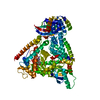
- Structure visualization
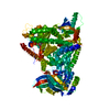
| Structure viewer | Molecule: MolmilJmol/JSmol |
|---|
- Downloads & links
-Download
| PDBx/mmCIF format | 1ff9.cif.gz | 100.8 KB | Display | PDBx/mmCIF format |
|---|---|---|---|---|
| PDB format | pdb1ff9.ent.gz | 77.3 KB | Display | PDB format |
| PDBx/mmJSON format | 1ff9.json.gz | Tree view | PDBx/mmJSON format | |
| Others |  Other downloads Other downloads |
-Validation report
| Summary document | 1ff9_validation.pdf.gz | 442.1 KB | Display | wwPDB validaton report |
|---|---|---|---|---|
| Full document | 1ff9_full_validation.pdf.gz | 457.4 KB | Display | |
| Data in XML | 1ff9_validation.xml.gz | 21.3 KB | Display | |
| Data in CIF | 1ff9_validation.cif.gz | 30 KB | Display | |
| Arichive directory | https://data.pdbj.org/pub/pdb/validation_reports/ff/1ff9ftp://data.pdbj.org/pub/pdb/validation_reports/ff/1ff9 | HTTPS FTP |
-Related structure data


-Links
 PDBj
PDBj- Assembly
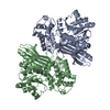
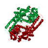
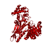
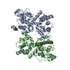
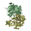
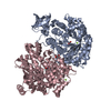
| Deposited unit | 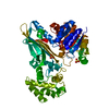
| ||||||||
|---|---|---|---|---|---|---|---|---|---|
| 1 | 
| ||||||||
| Unit cell |
|
-Components
| #1: Protein | Mass: 49156.262 Da / Num. of mol.: 1 Source method: isolated from a genetically manipulated source Source: (gene. exp.) Magnaporthe grisea (fungus) / Production host:  References: UniProt: Q9P4R4, saccharopine dehydrogenase (NAD+, L-lysine-forming) | ||
|---|---|---|---|
| #2: Chemical |   Mass: 96.063 Da / Num. of mol.: 3 / Source method: obtained synthetically / Formula: SO4 Mass: 96.063 Da / Num. of mol.: 3 / Source method: obtained synthetically / Formula: SO4#3: Water | ChemComp-HOH / |  Mass: 18.015 Da / Num. of mol.: 190 / Source method: isolated from a natural source / Formula: H2O Mass: 18.015 Da / Num. of mol.: 190 / Source method: isolated from a natural source / Formula: H2O |
-Experimental details
-Experiment
| Experiment | Method: X-RAY DIFFRACTION / Number of used crystals: 1 |
|---|
- Sample preparation
| Crystal | Density Matthews: 2.29 Å3/Da / Density % sol: 46.35 % | |||||||||||||||||||||||||
|---|---|---|---|---|---|---|---|---|---|---|---|---|---|---|---|---|---|---|---|---|---|---|---|---|---|---|
| Crystal grow | Temperature: 277 K / Method: vapor diffusion / pH: 4.8 Details: ammonium sulphate, sodium acetate, DTT, pH 4.8, VAPOR DIFFUSION, temperature 277K | |||||||||||||||||||||||||
| Crystal grow | *PLUS Temperature: 4 ℃ / Method: vapor diffusion, hanging drop | |||||||||||||||||||||||||
| Components of the solutions | *PLUS
|
-Data collection
| Diffraction | Mean temperature: 100 K |
|---|---|
| Diffraction source | Source: SYNCHROTRON / Site: EMBL/DESY, HAMBURG  / Beamline: X11 / Wavelength: 0.9058 / Beamline: X11 / Wavelength: 0.9058 |
| Detector | Type: MARRESEARCH / Detector: IMAGE PLATE / Date: May 19, 1998 |
| Radiation | Protocol: SINGLE WAVELENGTH / Monochromatic (M) / Laue (L): M / Scattering type: x-ray |
| Radiation wavelength | Wavelength: 0.9058 Å / Relative weight: 1 |
| Reflection | Resolution: 2→24.91 Å / Num. all: 29843 / Num. obs: 578167 / % possible obs: 98.5 % / Observed criterion σ(F): 0 / Observed criterion σ(I): -3 / Redundancy: 19.4 % / Biso Wilson estimate: 22.3 Å2 / Limit h max: 53 / Limit h min: -57 / Limit k max: 28 / Limit k min: -57 / Limit l max: 37 / Limit l min: 0 / Observed criterion F max: 1953730.55 / Observed criterion F min: 11.1 / Rmerge(I) obs: 0.053 / Net I/σ(I): 25.5 |
| Reflection shell | Resolution: 2→2.05 Å / Redundancy: 3.5 % / Rmerge(I) obs: 0.33 / Num. unique all: 1994 / % possible all: 98.3 |
| Reflection | *PLUS Num. obs: 29843 / Num. measured all: 578167 |
| Reflection shell | *PLUS % possible obs: 98.3 % / Rmerge(I) obs: 0.33 / Mean I/σ(I) obs: 3.3 |
- Processing
| Software |
| ||||||||||||||||||||||||||||||||||||||||||||||||||||||||||||
|---|---|---|---|---|---|---|---|---|---|---|---|---|---|---|---|---|---|---|---|---|---|---|---|---|---|---|---|---|---|---|---|---|---|---|---|---|---|---|---|---|---|---|---|---|---|---|---|---|---|---|---|---|---|---|---|---|---|---|---|---|---|
| Refinement | Resolution: 2→24.91 Å / Rfactor Rfree error: 0.007 / Occupancy max: 1.01 / Occupancy min: 1 / Cross valid method: THROUGHOUT / σ(F): 0 / σ(I): 0
| ||||||||||||||||||||||||||||||||||||||||||||||||||||||||||||
| Solvent computation | Solvent model: CNS bulk solvent model used / Bsol: 52.8425 Å2 / ksol: 0.347881 e/Å3 | ||||||||||||||||||||||||||||||||||||||||||||||||||||||||||||
| Displacement parameters | Biso max: 85.56 Å2 / Biso mean: 40.66 Å2 / Biso min: 19.69 Å2
| ||||||||||||||||||||||||||||||||||||||||||||||||||||||||||||
| Refine analyze |
| ||||||||||||||||||||||||||||||||||||||||||||||||||||||||||||
| Refinement step | Cycle: LAST / Resolution: 2→24.91 Å
| ||||||||||||||||||||||||||||||||||||||||||||||||||||||||||||
| Refine LS restraints |
| ||||||||||||||||||||||||||||||||||||||||||||||||||||||||||||
| LS refinement shell | Resolution: 2→2.09 Å / Rfactor Rfree error: 0.02
| ||||||||||||||||||||||||||||||||||||||||||||||||||||||||||||
| Xplor file |
| ||||||||||||||||||||||||||||||||||||||||||||||||||||||||||||
| Software | *PLUS Name: CNS / Classification: refinement | ||||||||||||||||||||||||||||||||||||||||||||||||||||||||||||
| Refinement | *PLUS σ(F): 0 / % reflection Rfree: 5.1 % / Rfactor obs: 0.241 | ||||||||||||||||||||||||||||||||||||||||||||||||||||||||||||
| Solvent computation | *PLUS | ||||||||||||||||||||||||||||||||||||||||||||||||||||||||||||
| Displacement parameters | *PLUS | ||||||||||||||||||||||||||||||||||||||||||||||||||||||||||||
| Refine LS restraints | *PLUS
| ||||||||||||||||||||||||||||||||||||||||||||||||||||||||||||
| LS refinement shell | *PLUS Rfactor Rfree: 0.29 / Rfactor Rwork: 0.288 / Rfactor obs: 0.283 |
