 ムービー
ムービー コントローラー


+ データを開く

- 基本情報
基本情報
| 登録情報 | データベース: PDB / ID: 1f8a | ||||||
|---|---|---|---|---|---|---|---|
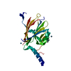
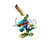
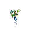
| タイトル | STRUCTURAL BASIS FOR THE PHOSPHOSERINE-PROLINE RECOGNITION BY GROUP IV WW DOMAINS | ||||||
 要素 要素 |
| ||||||
 キーワード キーワード | ISOMERASE / Peptidyl-Proline Isomerase / WW domain / phosphoserine binding | ||||||
| 機能・相同性 |  機能・相同性情報 機能・相同性情報cis-trans isomerase activity / phosphothreonine residue binding / negative regulation of cell motility / ubiquitin ligase activator activity / regulation of protein localization to nucleus / GTPase activating protein binding / protein targeting to mitochondrion / protein peptidyl-prolyl isomerization / mitogen-activated protein kinase kinase binding / regulation of mitotic nuclear division ...cis-trans isomerase activity / phosphothreonine residue binding / negative regulation of cell motility / ubiquitin ligase activator activity / regulation of protein localization to nucleus / GTPase activating protein binding / protein targeting to mitochondrion / protein peptidyl-prolyl isomerization / mitogen-activated protein kinase kinase binding / regulation of mitotic nuclear division / negative regulation of SMAD protein signal transduction / PI5P Regulates TP53 Acetylation / negative regulation of amyloid-beta formation / cytoskeletal motor activity / postsynaptic cytosol / RHO GTPases Activate NADPH Oxidases / phosphoserine residue binding / negative regulation of protein binding / positive regulation of GTPase activity / peptidyl-prolyl cis-trans isomerase activity / regulation of cytokinesis / RNA polymerase II CTD heptapeptide repeat P3 isomerase activity / RNA polymerase II CTD heptapeptide repeat P6 isomerase activity / peptidylprolyl isomerase / phosphoprotein binding / Negative regulators of DDX58/IFIH1 signaling / negative regulation of transforming growth factor beta receptor signaling pathway / synapse organization / negative regulation of ERK1 and ERK2 cascade / regulation of protein stability / negative regulation of protein catabolic process / beta-catenin binding / tau protein binding / ISG15 antiviral mechanism / neuron differentiation / positive regulation of canonical Wnt signaling pathway / positive regulation of protein phosphorylation / regulation of gene expression / midbody / cellular response to hypoxia / Regulation of TP53 Activity through Phosphorylation / response to hypoxia / protein stabilization / nuclear speck / ciliary basal body / glutamatergic synapse / positive regulation of transcription by RNA polymerase II / nucleoplasm / nucleus / cytosol / cytoplasm 類似検索 - 分子機能 | ||||||
| 生物種 |  Homo sapiens (ヒト) Homo sapiens (ヒト) | ||||||
| 手法 |  X線回折 / シンクロトロン / 解像度: 1.84 Å X線回折 / シンクロトロン / 解像度: 1.84 Å | ||||||
 データ登録者 データ登録者 | Verdecia, M.A. / Bowman, M.E. / Lu, K.P. / Hunter, T. / Noel, J.P. | ||||||
 引用 引用 | ジャーナル: Nat.Struct.Biol. / 年: 2000 タイトル: Structural basis for phosphoserine-proline recognition by group IV WW domains. 著者: Verdecia, M.A. / Bowman, M.E. / Lu, K.P. / Hunter, T. / Noel, J.P. | ||||||
| 履歴 |
|
- 構造の表示
構造の表示
| 構造ビューア | 分子: MolmilJmol/JSmol |
|---|
- ダウンロードとリンク
-ダウンロード
| PDBx/mmCIF形式 | 1f8a.cif.gz | 49 KB | 表示 | PDBx/mmCIF形式 |
|---|---|---|---|---|
| PDB形式 | pdb1f8a.ent.gz | 34.1 KB | 表示 | PDB形式 |
| PDBx/mmJSON形式 | 1f8a.json.gz | ツリー表示 | PDBx/mmJSON形式 | |
| その他 |  その他のダウンロード その他のダウンロード |
-検証レポート
| アーカイブディレクトリ | https://data.pdbj.org/pub/pdb/validation_reports/f8/1f8aftp://data.pdbj.org/pub/pdb/validation_reports/f8/1f8a | HTTPS FTP |
|---|
-関連構造データ


-リンク
 PDBj
PDBj


- 集合体
集合体
| 登録構造単位 | 
| ||||||||
|---|---|---|---|---|---|---|---|---|---|
| 1 |
| ||||||||
| 単位格子 |
| ||||||||
| 詳細 | The biological assembly is a monomer of Pin1 bound to the phosphorylated peptide |
-要素
| #1: タンパク質 | 分子量: 18610.641 Da / 分子数: 1 / 由来タイプ: 組換発現 / 由来: (組換発現) Homo sapiens (ヒト) / 発現宿主:  |
|---|---|
| #2: タンパク質・ペプチド | 分子量: 897.714 Da / 分子数: 1 / 由来タイプ: 合成 / 詳細: SOLID-PHASE PEPTIDE SYNTHESIS |
| #3: 水 | ChemComp-HOH /  分子量: 18.015 Da / 分子数: 152 / 由来タイプ: 天然 / 式: H2O 分子量: 18.015 Da / 分子数: 152 / 由来タイプ: 天然 / 式: H2O |
| Has protein modification | Y |
-実験情報
-実験
| 実験 | 手法: X線回折 / 使用した結晶の数: 1 |
|---|
- 試料調製
試料調製
| 結晶 | マシュー密度: 2.47 Å3/Da / 溶媒含有率: 50.25 % | ||||||||||||||||||||
|---|---|---|---|---|---|---|---|---|---|---|---|---|---|---|---|---|---|---|---|---|---|
| 結晶化 | 温度: 298 K / 手法: 蒸気拡散法, ハンギングドロップ法 / pH: 7 詳細: 100 mM MOPSO-Na+, 28% PEG 8000, 2 mM DTT, pH 7.0, VAPOR DIFFUSION, HANGING DROP, temperature 298.0K | ||||||||||||||||||||
| 結晶化 | *PLUS 温度: 4 ℃ | ||||||||||||||||||||
| 溶液の組成 | *PLUS
|
-データ収集
| 回折 | 平均測定温度: 100 K |
|---|---|
| 放射光源 | 由来: シンクロトロン / サイト: SSRL  / ビームライン: BL9-1 / 波長: 0.98 / ビームライン: BL9-1 / 波長: 0.98 |
| 検出器 | タイプ: MACSCIENCE DIP100S / 検出器: IMAGE PLATE / 日付: 1999年4月15日 |
| 放射 | プロトコル: SINGLE WAVELENGTH / 単色(M)・ラウエ(L): M / 散乱光タイプ: x-ray |
| 放射波長 | 波長: 0.98 Å / 相対比: 1 |
| 反射 | 解像度: 1.84→62.02 Å / Num. obs: 293095 / % possible obs: 97.3 % / Observed criterion σ(I): 2 / Biso Wilson estimate: 17.4 Å2 / Rmerge(I) obs: 0.062 / Net I/σ(I): 21.5 |
| 反射 シェル | 解像度: 1.84→1.9 Å / Rmerge(I) obs: 0.337 / Num. unique all: 17107 / % possible all: 98.2 |
| 反射 | *PLUS Num. obs: 17107 / Num. measured all: 293095 |
| 反射 シェル | *PLUS % possible obs: 98.2 % / Mean I/σ(I) obs: 4.3 |
- 解析
解析
| ソフトウェア |
| ||||||||||||||||||||||||||||||||||||||||||||
|---|---|---|---|---|---|---|---|---|---|---|---|---|---|---|---|---|---|---|---|---|---|---|---|---|---|---|---|---|---|---|---|---|---|---|---|---|---|---|---|---|---|---|---|---|---|
| 精密化 | 解像度: 1.84→41.41 Å / Rfactor Rfree error: 0.009 / Data cutoff high absF: 1085629.02 / Data cutoff low absF: 0 / Isotropic thermal model: RESTRAINED / 交差検証法: THROUGHOUT / σ(F): 0 / σ(I): 2 / 立体化学のターゲット値: Engh & Huber
| ||||||||||||||||||||||||||||||||||||||||||||
| 溶媒の処理 | 溶媒モデル: FLAT MODEL / Bsol: 36 Å2 / ksol: 0.3734 e/Å3 | ||||||||||||||||||||||||||||||||||||||||||||
| 原子変位パラメータ | Biso mean: 27.3 Å2
| ||||||||||||||||||||||||||||||||||||||||||||
| Refine analyze |
| ||||||||||||||||||||||||||||||||||||||||||||
| 精密化ステップ | サイクル: LAST / 解像度: 1.84→41.41 Å
| ||||||||||||||||||||||||||||||||||||||||||||
| 拘束条件 |
| ||||||||||||||||||||||||||||||||||||||||||||
| LS精密化 シェル | 解像度: 1.84→1.96 Å / Rfactor Rfree error: 0.024 / Total num. of bins used: 6
| ||||||||||||||||||||||||||||||||||||||||||||
| Xplor file |
| ||||||||||||||||||||||||||||||||||||||||||||
| ソフトウェア | *PLUS 名称: CNS / バージョン: 1 / 分類: refinement | ||||||||||||||||||||||||||||||||||||||||||||
| 精密化 | *PLUS σ(F): 0 / % reflection Rfree: 5.1 % | ||||||||||||||||||||||||||||||||||||||||||||
| 溶媒の処理 | *PLUS | ||||||||||||||||||||||||||||||||||||||||||||
| 原子変位パラメータ | *PLUS Biso mean: 27.3 Å2 | ||||||||||||||||||||||||||||||||||||||||||||
| 拘束条件 | *PLUS
| ||||||||||||||||||||||||||||||||||||||||||||
| LS精密化 シェル | *PLUS Rfactor Rfree: 0.29 / % reflection Rfree: 5.4 % / Rfactor Rwork: 0.239 |
