 Movie
Movie Controller

[English] 日本語
 Yorodumi
- PDB-1f7v: CRYSTAL STRUCTURE OF YEAST ARGINYL-TRNA SYNTHETASE COMPLEXED WITH... -
+ Open data

- Basic information
| Entry | Database: PDB / ID: 1f7v | ||||||
|---|---|---|---|---|---|---|---|
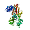
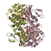
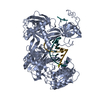
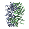
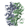
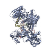
| Title | CRYSTAL STRUCTURE OF YEAST ARGINYL-TRNA SYNTHETASE COMPLEXED WITH THE TRNAARG | ||||||
 Components Components |
| ||||||
 Keywords Keywords | LIGASE/RNA / tRNA-protein complex / aminoacylation / arginyl-tRNA synthetase / LIGASE-RNA COMPLEX | ||||||
| Function / homology |  Function and homology information Function and homology informationarginine-tRNA ligase / arginine-tRNA ligase activity / arginyl-tRNA aminoacylation / sequence-specific mRNA binding / mitochondrial translation / mitochondrion / ATP binding / cytosol Similarity search - Function | ||||||
| Biological species |  | ||||||
| Method |  X-RAY DIFFRACTION / SYNCHROTRON / Resolution: 2.9 Å X-RAY DIFFRACTION / SYNCHROTRON / Resolution: 2.9 Å | ||||||
 Authors Authors | Delagoutte, B. / Moras, D. / Cavarelli, J. | ||||||
 Citation Citation | Journal: EMBO J. / Year: 2000 Title: tRNA aminoacylation by arginyl-tRNA synthetase: induced conformations during substrates binding Authors: Delagoutte, B. / Moras, D. / Cavarelli, J. #1: Journal: Acta Crystallogr.,Sect.D / Year: 2000Title: Crystallization and preliminary X-ray crystallographic analysis of yeast arginyl-tRNA synthetase - yeast tRNAArg complexes Authors: Delagoutte, B. / Keith, G. / Moras, D. / Cavarelli, J. | ||||||
| History |
|
- Structure visualization
| Structure viewer | Molecule: MolmilJmol/JSmol |
|---|
- Downloads & links
-Download
| PDBx/mmCIF format | 1f7v.cif.gz | 179.2 KB | Display | PDBx/mmCIF format |
|---|---|---|---|---|
| PDB format | pdb1f7v.ent.gz | 136.3 KB | Display | PDB format |
| PDBx/mmJSON format | 1f7v.json.gz | Tree view | PDBx/mmJSON format | |
| Others |  Other downloads Other downloads |
-Validation report
| Arichive directory | https://data.pdbj.org/pub/pdb/validation_reports/f7/1f7vftp://data.pdbj.org/pub/pdb/validation_reports/f7/1f7v | HTTPS FTP |
|---|
-Related structure data


-Links
 PDBj
PDBj


- Assembly
| Deposited unit | 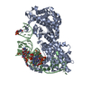
| ||||||||||
|---|---|---|---|---|---|---|---|---|---|---|---|
| 1 |
| ||||||||||
| Unit cell |
|
-Components
| #1: RNA chain | Mass: 24573.770 Da / Num. of mol.: 1 / Source method: obtained synthetically Details: this sequence occurs naturally in yeast Saccharomyces cerevisae |
|---|---|
| #2: Protein | Mass: 69617.867 Da / Num. of mol.: 1 Source method: isolated from a genetically manipulated source Source: (gene. exp.) Plasmid: PTRC99 / Production host:  |
| #3: Water | ChemComp-HOH /  Mass: 18.015 Da / Num. of mol.: 44 / Source method: isolated from a natural source / Formula: H2O Mass: 18.015 Da / Num. of mol.: 44 / Source method: isolated from a natural source / Formula: H2O |
-Experimental details
-Experiment
| Experiment | Method: X-RAY DIFFRACTION / Number of used crystals: 1 |
|---|
- Sample preparation
| Crystal | Density Matthews: 3.41 Å3/Da / Density % sol: 63.91 % | ||||||||||||||||||||||||||||||||||||||||||||||||
|---|---|---|---|---|---|---|---|---|---|---|---|---|---|---|---|---|---|---|---|---|---|---|---|---|---|---|---|---|---|---|---|---|---|---|---|---|---|---|---|---|---|---|---|---|---|---|---|---|---|
| Crystal grow | Temperature: 277 K / Method: vapor diffusion, hanging drop / pH: 6.5 Details: 2.1 M (NH4)2SO4, hexanediol, pH 6.5, VAPOR DIFFUSION, HANGING DROP at 277K | ||||||||||||||||||||||||||||||||||||||||||||||||
| Components of the solutions |
| ||||||||||||||||||||||||||||||||||||||||||||||||
| Crystal grow | *PLUS Details: Delagoutte, B., (2000) Acta Crystallogr., Sect.D, 56, 492. | ||||||||||||||||||||||||||||||||||||||||||||||||
| Components of the solutions | *PLUS
|
-Data collection
| Diffraction | Mean temperature: 110 K |
|---|---|
| Diffraction source | Source: SYNCHROTRON / Site: ESRF  / Beamline: ID14-3 / Wavelength: 0.931 / Beamline: ID14-3 / Wavelength: 0.931 |
| Detector | Type: ADSC QUANTUM 4 / Detector: CCD / Date: Nov 25, 1999 |
| Radiation | Protocol: SINGLE WAVELENGTH / Monochromatic (M) / Laue (L): M / Scattering type: x-ray |
| Radiation wavelength | Wavelength: 0.931 Å / Relative weight: 1 |
| Reflection | Resolution: 2.9→25 Å / Num. all: 28856 / Num. obs: 28856 / % possible obs: 100 % / Observed criterion σ(F): 0 / Observed criterion σ(I): 0 / Redundancy: 7 % / Biso Wilson estimate: 56.9 Å2 / Rmerge(I) obs: 0.056 / Net I/σ(I): 30.2 |
| Reflection shell | Resolution: 2.9→3 Å / Redundancy: 6.9 % / Rmerge(I) obs: 0.262 / Num. unique all: 2835 / % possible all: 100 |
| Reflection | *PLUS |
- Processing
| Software |
| ||||||||||||||||||||||||||||||||||||||||||||||||||||||||||||||||||||||||||||||||
|---|---|---|---|---|---|---|---|---|---|---|---|---|---|---|---|---|---|---|---|---|---|---|---|---|---|---|---|---|---|---|---|---|---|---|---|---|---|---|---|---|---|---|---|---|---|---|---|---|---|---|---|---|---|---|---|---|---|---|---|---|---|---|---|---|---|---|---|---|---|---|---|---|---|---|---|---|---|---|---|---|---|
| Refinement | Resolution: 2.9→24.87 Å / Rfactor Rfree error: 0.005 / Data cutoff high absF: 2531107.58 / Data cutoff low absF: 0 / Isotropic thermal model: RESTRAINED / Cross valid method: THROUGHOUT / σ(F): 0 / σ(I): 0 / Stereochemistry target values: Engh & Huber
| ||||||||||||||||||||||||||||||||||||||||||||||||||||||||||||||||||||||||||||||||
| Solvent computation | Solvent model: FLAT MODEL / Bsol: 26.52 Å2 / ksol: 0.338 e/Å3 | ||||||||||||||||||||||||||||||||||||||||||||||||||||||||||||||||||||||||||||||||
| Displacement parameters | Biso mean: 39.4 Å2
| ||||||||||||||||||||||||||||||||||||||||||||||||||||||||||||||||||||||||||||||||
| Refine analyze |
| ||||||||||||||||||||||||||||||||||||||||||||||||||||||||||||||||||||||||||||||||
| Refinement step | Cycle: LAST / Resolution: 2.9→24.87 Å
| ||||||||||||||||||||||||||||||||||||||||||||||||||||||||||||||||||||||||||||||||
| Refine LS restraints |
| ||||||||||||||||||||||||||||||||||||||||||||||||||||||||||||||||||||||||||||||||
| LS refinement shell | Resolution: 2.9→3.08 Å / Rfactor Rfree error: 0.016 / Total num. of bins used: 6
| ||||||||||||||||||||||||||||||||||||||||||||||||||||||||||||||||||||||||||||||||
| Software | *PLUS Name: CNS / Version: 1 / Classification: refinement | ||||||||||||||||||||||||||||||||||||||||||||||||||||||||||||||||||||||||||||||||
| Refinement | *PLUS σ(F): 0 / % reflection Rfree: 10 % | ||||||||||||||||||||||||||||||||||||||||||||||||||||||||||||||||||||||||||||||||
| Solvent computation | *PLUS | ||||||||||||||||||||||||||||||||||||||||||||||||||||||||||||||||||||||||||||||||
| Displacement parameters | *PLUS Biso mean: 39.4 Å2 | ||||||||||||||||||||||||||||||||||||||||||||||||||||||||||||||||||||||||||||||||
| Refine LS restraints | *PLUS
| ||||||||||||||||||||||||||||||||||||||||||||||||||||||||||||||||||||||||||||||||
| LS refinement shell | *PLUS Rfactor Rfree: 0.36 / % reflection Rfree: 10.5 % / Rfactor Rwork: 0.295 |
