 Movie
Movie Controller

[English] 日本語
 Yorodumi
- PDB-1f75: CRYSTAL STRUCTURE OF UNDECAPRENYL DIPHOSPHATE SYNTHASE FROM MICRO... -
+ Open data

- Basic information
| Entry | Database: PDB / ID: 1f75 | ||||||
|---|---|---|---|---|---|---|---|
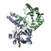
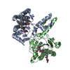
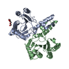
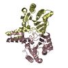
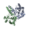
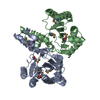
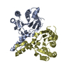
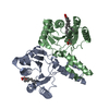
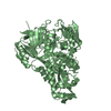
| Title | CRYSTAL STRUCTURE OF UNDECAPRENYL DIPHOSPHATE SYNTHASE FROM MICROCOCCUS LUTEUS B-P 26 | ||||||
 Components Components | UNDECAPRENYL PYROPHOSPHATE SYNTHETASE | ||||||
 Keywords Keywords | TRANSFERASE / PARALLEL BETA SHEET / new fold for Isoprenoid Synthase / PEPTIDOGLYCAN SYNTHESIS | ||||||
| Function / homology |  Function and homology information Function and homology informationditrans,polycis-undecaprenyl-diphosphate synthase [(2E,6E)-farnesyl-diphosphate specific] / ditrans,polycis-undecaprenyl-diphosphate synthase [(2E,6E)-farnesyl-diphosphate specific] activity / polyprenol biosynthetic process / manganese ion binding / magnesium ion binding / cytosol Similarity search - Function | ||||||
| Biological species |  Micrococcus luteus (bacteria) Micrococcus luteus (bacteria) | ||||||
| Method |  X-RAY DIFFRACTION / SYNCHROTRON / MIRAS / Resolution: 2.2 Å X-RAY DIFFRACTION / SYNCHROTRON / MIRAS / Resolution: 2.2 Å | ||||||
 Authors Authors | Fujihashi, M. / Zhang, Y.-W. / Higuchi, Y. / Li, X.-Y. / Koyama, T. / Miki, K. | ||||||
 Citation Citation | Journal: Proc.Natl.Acad.Sci.USA / Year: 2001 Title: Crystal structure of cis-prenyl chain elongating enzyme, undecaprenyl diphosphate synthase. Authors: Fujihashi, M. / Zhang, Y.W. / Higuchi, Y. / Li, X.Y. / Koyama, T. / Miki, K. #1: Journal: Acta Crystallogr.,Sect.D / Year: 1999Title: Crystallization and preliminary X-ray diffraction studies of undecaprenyl diphosphate synthase from Micrococcus luteus B-P 26 Authors: Fujihashi, M. / Shimizu, N. / Zhang, Y.W. / Koyama, T. / Miki, K. | ||||||
| History |
|
- Structure visualization
| Structure viewer | Molecule: MolmilJmol/JSmol |
|---|
- Downloads & links
-Download
| PDBx/mmCIF format | 1f75.cif.gz | 97.5 KB | Display | PDBx/mmCIF format |
|---|---|---|---|---|
| PDB format | pdb1f75.ent.gz | 75.6 KB | Display | PDB format |
| PDBx/mmJSON format | 1f75.json.gz | Tree view | PDBx/mmJSON format | |
| Others |  Other downloads Other downloads |
-Validation report
| Arichive directory | https://data.pdbj.org/pub/pdb/validation_reports/f7/1f75ftp://data.pdbj.org/pub/pdb/validation_reports/f7/1f75 | HTTPS FTP |
|---|
-Related structure data
| Similar structure data |
|---|

-Links
 PDBj
PDBj- Assembly
| Deposited unit | 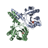
| ||||||||
|---|---|---|---|---|---|---|---|---|---|
| 1 |
| ||||||||
| Unit cell |
|
-Components
| #1: Protein | Mass: 28945.227 Da / Num. of mol.: 2 / Mutation: K54R Source method: isolated from a genetically manipulated source Source: (gene. exp.) Micrococcus luteus (bacteria) / Plasmid: PMLUEX3 / Production host: References: UniProt: O82827, ditrans,polycis-undecaprenyl-diphosphate synthase [(2E,6E)-farnesyl-diphosphate specific] #2: Chemical |   Mass: 96.063 Da / Num. of mol.: 2 / Source method: obtained synthetically / Formula: SO4 Mass: 96.063 Da / Num. of mol.: 2 / Source method: obtained synthetically / Formula: SO4#3: Water | ChemComp-HOH / |  Mass: 18.015 Da / Num. of mol.: 38 / Source method: isolated from a natural source / Formula: H2O Mass: 18.015 Da / Num. of mol.: 38 / Source method: isolated from a natural source / Formula: H2O |
|---|
-Experimental details
-Experiment
| Experiment | Method: X-RAY DIFFRACTION / Number of used crystals: 1 |
|---|
- Sample preparation
| Crystal | Density Matthews: 2.4 Å3/Da / Density % sol: 48.8 % | ||||||||||||||||||||||||||||||||||||||||||||||||
|---|---|---|---|---|---|---|---|---|---|---|---|---|---|---|---|---|---|---|---|---|---|---|---|---|---|---|---|---|---|---|---|---|---|---|---|---|---|---|---|---|---|---|---|---|---|---|---|---|---|
| Crystal grow | Temperature: 293 K / Method: vapor diffusion / pH: 5.4 Details: ammonium sulfate, lithium sulfate, pH 5.4, VAPOR DIFFUSION, temperature 293.0K | ||||||||||||||||||||||||||||||||||||||||||||||||
| Crystal grow | *PLUS pH: 8 / Method: vapor diffusion, sitting dropDetails: drop consists of equal amounts of protein and reservoir solutions | ||||||||||||||||||||||||||||||||||||||||||||||||
| Components of the solutions | *PLUS
|
-Data collection
| Diffraction | Mean temperature: 293 K |
|---|---|
| Diffraction source | Source: SYNCHROTRON / Site: Photon Factory  / Beamline: BL-6B / Wavelength: 1 / Beamline: BL-6B / Wavelength: 1 |
| Detector | Type: RIGAKU / Detector: IMAGE PLATE / Date: Feb 4, 1999 |
| Radiation | Protocol: SINGLE WAVELENGTH / Monochromatic (M) / Laue (L): M / Scattering type: x-ray |
| Radiation wavelength | Wavelength: 1 Å / Relative weight: 1 |
| Reflection | Resolution: 2.2→100 Å / Num. all: 10 / Num. obs: 57323 / % possible obs: 83.7 % / Observed criterion σ(I): 1 / Redundancy: 2.43 % / Biso Wilson estimate: 30.1 Å2 / Rmerge(I) obs: 0.032 / Net I/σ(I): 24.3 |
| Reflection shell | Resolution: 2.2→2.24 Å / Rmerge(I) obs: 0.168 / % possible all: 56.8 |
| Reflection | *PLUS Num. obs: 23530 / Num. measured all: 57323 |
| Reflection shell | *PLUS % possible obs: 56.8 % |
- Processing
| Software |
| ||||||||||||||||||||||||||||||
|---|---|---|---|---|---|---|---|---|---|---|---|---|---|---|---|---|---|---|---|---|---|---|---|---|---|---|---|---|---|---|---|
| Refinement | Method to determine structure: MIRAS / Resolution: 2.2→50 Å / σ(F): 2 / Stereochemistry target values: Engh & Huber
| ||||||||||||||||||||||||||||||
| Refinement step | Cycle: LAST / Resolution: 2.2→50 Å
| ||||||||||||||||||||||||||||||
| Software | *PLUS Name: X-PLOR / Version: 3.843 / Classification: refinement | ||||||||||||||||||||||||||||||
| Refine LS restraints | *PLUS
|
