 Movie
Movie Controller

[English] 日本語
 Yorodumi
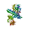
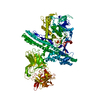
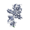
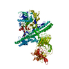
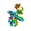
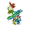
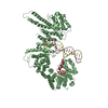
- PDB-1f31: CRYSTAL STRUCTURE OF CLOSTRIDIUM BOTULINUM NEUROTOXIN B COMPLEXED... -
+ Open data

- Basic information
| Entry | Database: PDB / ID: 1f31 | |||||||||
|---|---|---|---|---|---|---|---|---|---|---|
| Title | CRYSTAL STRUCTURE OF CLOSTRIDIUM BOTULINUM NEUROTOXIN B COMPLEXED WITH A TRISACCHARIDE | |||||||||
 Components Components | BOTULINUM NEUROTOXIN TYPE B | |||||||||
 Keywords Keywords | TOXIN / botulinum / zinc / metalloprotease / transmembrane / neurotoxin / complex / ganglioside | |||||||||
| Function / homology |  Function and homology information Function and homology informationToxicity of botulinum toxin type B (botB) / bontoxilysin / host cell presynaptic membrane / host cell cytoplasmic vesicle / host cell cytosol / protein transmembrane transporter activity / metalloendopeptidase activity / toxin activity / lipid binding / host cell plasma membrane ...Toxicity of botulinum toxin type B (botB) / bontoxilysin / host cell presynaptic membrane / host cell cytoplasmic vesicle / host cell cytosol / protein transmembrane transporter activity / metalloendopeptidase activity / toxin activity / lipid binding / host cell plasma membrane / proteolysis / extracellular region / zinc ion binding / membrane Similarity search - Function | |||||||||
| Biological species |   Clostridium botulinum (bacteria) Clostridium botulinum (bacteria) | |||||||||
| Method |  X-RAY DIFFRACTION / SYNCHROTRON / Resolution: 2.6 Å X-RAY DIFFRACTION / SYNCHROTRON / Resolution: 2.6 Å | |||||||||
 Authors Authors | Swaminathan, S. / Eswaramoorthy, S. | |||||||||
 Citation Citation | Journal: Nat.Struct.Biol. / Year: 2000 Title: Structural analysis of the catalytic and binding sites of Clostridium botulinum neurotoxin B. Authors: Swaminathan, S. / Eswaramoorthy, S. #1: Journal: Acta Crystallogr.,Sect.D / Year: 2000Title: Crystallization and Preliminary X-ray Analysis of Clostridium botulinum Neurotoxin Type B Authors: Swaminathan, S. / Eswaramoorthy, S. | |||||||||
| History |
|
- Structure visualization
| Structure viewer | Molecule: MolmilJmol/JSmol |
|---|
- Downloads & links
-Download
| PDBx/mmCIF format | 1f31.cif.gz | 275.1 KB | Display | PDBx/mmCIF format |
|---|---|---|---|---|
| PDB format | pdb1f31.ent.gz | 215.3 KB | Display | PDB format |
| PDBx/mmJSON format | 1f31.json.gz | Tree view | PDBx/mmJSON format | |
| Others |  Other downloads Other downloads |
-Validation report
| Summary document | 1f31_validation.pdf.gz | 471 KB | Display | wwPDB validaton report |
|---|---|---|---|---|
| Full document | 1f31_full_validation.pdf.gz | 496.1 KB | Display | |
| Data in XML | 1f31_validation.xml.gz | 28.3 KB | Display | |
| Data in CIF | 1f31_validation.cif.gz | 43.3 KB | Display | |
| Arichive directory | https://data.pdbj.org/pub/pdb/validation_reports/f3/1f31ftp://data.pdbj.org/pub/pdb/validation_reports/f3/1f31 | HTTPS FTP |
-Related structure data
| Related structure data |  1epwSC S: Starting model for refinement C: citing same article ( |
|---|---|
| Similar structure data |


-Links
 PDBj
PDBj- Assembly
| Deposited unit | 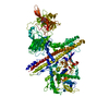
| ||||||||
|---|---|---|---|---|---|---|---|---|---|
| 1 |
| ||||||||
| Unit cell |
|
-Components
| #1: Protein | Mass: 150833.375 Da / Num. of mol.: 1 / Source method: isolated from a natural source / Source: (natural) Clostridium botulinum (bacteria) / References: UniProt: P10844, bontoxilysin |
|---|---|
| #2: Polysaccharide | N-acetyl-alpha-neuraminic acid-(2-3)-alpha-D-galactopyranose-(1-4)-alpha-D-glucopyranose Source method: isolated from a genetically manipulated source |
| #3: Chemical | ChemComp-ZN /   Mass: 65.409 Da / Num. of mol.: 1 / Source method: obtained synthetically / Formula: Zn Mass: 65.409 Da / Num. of mol.: 1 / Source method: obtained synthetically / Formula: Zn |
| #4: Chemical | ChemComp-SO4 /   Mass: 96.063 Da / Num. of mol.: 1 / Source method: obtained synthetically / Formula: SO4 Mass: 96.063 Da / Num. of mol.: 1 / Source method: obtained synthetically / Formula: SO4 |
| #5: Water | ChemComp-HOH /  Mass: 18.015 Da / Num. of mol.: 98 / Source method: isolated from a natural source / Formula: H2O Mass: 18.015 Da / Num. of mol.: 98 / Source method: isolated from a natural source / Formula: H2O |
| Has protein modification | Y |
-Experimental details
-Experiment
| Experiment | Method: X-RAY DIFFRACTION / Number of used crystals: 1 |
|---|
- Sample preparation
| Crystal | Density Matthews: 2.75 Å3/Da / Density % sol: 55 % | |||||||||||||||
|---|---|---|---|---|---|---|---|---|---|---|---|---|---|---|---|---|
| Crystal grow | Temperature: 291 K / Method: vapor diffusion, sitting drop / pH: 6 Details: PEG 4000, MES, pH 6.0, VAPOR DIFFUSION, SITTING DROP, temperature 291.0K | |||||||||||||||
| Crystal grow | *PLUS Method: unknown | |||||||||||||||
| Components of the solutions | *PLUS
|
-Data collection
| Diffraction | Mean temperature: 100 K |
|---|---|
| Diffraction source | Source: SYNCHROTRON / Site: NSLS  / Beamline: X25 / Wavelength: 1.1 / Beamline: X25 / Wavelength: 1.1 |
| Detector | Type: BRANDEIS - B4 / Detector: CCD / Date: Apr 21, 2000 |
| Radiation | Protocol: SINGLE WAVELENGTH / Monochromatic (M) / Laue (L): M / Scattering type: x-ray |
| Radiation wavelength | Wavelength: 1.1 Å / Relative weight: 1 |
| Reflection | Resolution: 2.3→50 Å / Num. all: 47458 / Num. obs: 47458 / % possible obs: 67 % / Redundancy: 2.2 % / Rmerge(I) obs: 0.052 |
| Reflection shell | Resolution: 2.3→2.38 Å / Rmerge(I) obs: 0.34 / % possible all: 12.9 |
| Reflection | *PLUS |
- Processing
| Software |
| |||||||||||||||||||||||||
|---|---|---|---|---|---|---|---|---|---|---|---|---|---|---|---|---|---|---|---|---|---|---|---|---|---|---|
| Refinement | Starting model: 1EPW Resolution: 2.6→50 Å / Cross valid method: FREE-R / σ(F): 2 / Stereochemistry target values: Engh & Huber
| |||||||||||||||||||||||||
| Refinement step | Cycle: LAST / Resolution: 2.6→50 Å
| |||||||||||||||||||||||||
| Refine LS restraints |
| |||||||||||||||||||||||||
| Software | *PLUS Name: CNS / Classification: refinement | |||||||||||||||||||||||||
| Refinement | *PLUS Highest resolution: 2.6 Å / Lowest resolution: 50 Å / σ(F): 2 / % reflection Rfree: 4 % / Rfactor all: 0.214 / Rfactor obs: 0.21 / Rfactor Rfree: 0.27 | |||||||||||||||||||||||||
| Solvent computation | *PLUS | |||||||||||||||||||||||||
| Displacement parameters | *PLUS Biso mean: 19.3 Å2 | |||||||||||||||||||||||||
| Refine LS restraints | *PLUS
|
