 Movie
Movie Controller

[English] 日本語
 Yorodumi
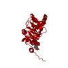
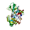
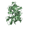
- PDB-1f2v: CRYSTAL STRUCTURE ANALYSIS OF PRECORRIN-8X METHYLMUTASE OF AEROBI... -
+ Open data

- Basic information
| Entry | Database: PDB / ID: 1f2v | ||||||
|---|---|---|---|---|---|---|---|
| Title | CRYSTAL STRUCTURE ANALYSIS OF PRECORRIN-8X METHYLMUTASE OF AEROBIC VITAMIN B12 SYNTHESIS | ||||||
 Components Components | PRECORRIN-8X METHYLMUTASE | ||||||
 Keywords Keywords | ISOMERASE / alpha-beta wind / doubly wound sheet | ||||||
| Function / homology |  Function and homology information Function and homology informationprecorrin-8X methylmutase / precorrin-8X methylmutase activity / cobalamin biosynthetic process Similarity search - Function | ||||||
| Biological species |  Pseudomonas denitrificans (bacteria) Pseudomonas denitrificans (bacteria) | ||||||
| Method |  X-RAY DIFFRACTION / Resolution: 2.1 Å X-RAY DIFFRACTION / Resolution: 2.1 Å | ||||||
 Authors Authors | Shipman, L.W. / Li, D. / Roessner, C.A. / Scott, A.I. / Sacchettini, J.C. | ||||||
 Citation Citation | Journal: Structure / Year: 2001 Title: Crystal structure of precorrin-8x methyl mutase. Authors: Shipman, L.W. / Li, D. / Roessner, C.A. / Scott, A.I. / Sacchettini, J.C. | ||||||
| History |
|
- Structure visualization
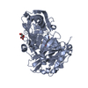
| Structure viewer | Molecule: MolmilJmol/JSmol |
|---|
- Downloads & links
-Download
| PDBx/mmCIF format | 1f2v.cif.gz | 55.2 KB | Display | PDBx/mmCIF format |
|---|---|---|---|---|
| PDB format | pdb1f2v.ent.gz | 39.4 KB | Display | PDB format |
| PDBx/mmJSON format | 1f2v.json.gz | Tree view | PDBx/mmJSON format | |
| Others |  Other downloads Other downloads |
-Validation report
| Arichive directory | https://data.pdbj.org/pub/pdb/validation_reports/f2/1f2vftp://data.pdbj.org/pub/pdb/validation_reports/f2/1f2v | HTTPS FTP |
|---|
-Related structure data

-Links
 PDBj
PDBj- Assembly
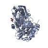
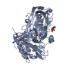
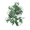
| Deposited unit | 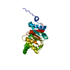
| ||||||||
|---|---|---|---|---|---|---|---|---|---|
| 1 | 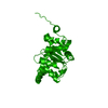
| ||||||||
| Unit cell |
| ||||||||
| Details | The biological assembly is a dimer constructed from chain A and symmetry partner generated by the two-fold. |
-Components
| #1: Protein | Mass: 23217.508 Da / Num. of mol.: 1 Source method: isolated from a genetically manipulated source Source: (gene. exp.) Pseudomonas denitrificans (bacteria) / Plasmid: PLM1 / Production host: |
|---|---|
| #2: Water | ChemComp-HOH /  Mass: 18.015 Da / Num. of mol.: 220 / Source method: isolated from a natural source / Formula: H2O Mass: 18.015 Da / Num. of mol.: 220 / Source method: isolated from a natural source / Formula: H2O |
-Experimental details
-Experiment
| Experiment | Method: X-RAY DIFFRACTION / Number of used crystals: 3 |
|---|
- Sample preparation
| Crystal | Density Matthews: 1.96 Å3/Da / Density % sol: 37.18 % | ||||||||||||||||||||||||||||||
|---|---|---|---|---|---|---|---|---|---|---|---|---|---|---|---|---|---|---|---|---|---|---|---|---|---|---|---|---|---|---|---|
| Crystal grow | Temperature: 289 K / Method: vapor diffusion, hanging drop / pH: 8.5 Details: PEG 4000, magnesium chloride, Tris-HCL, pH 8.5, VAPOR DIFFUSION, HANGING DROP, temperature 289K | ||||||||||||||||||||||||||||||
| Crystal grow | *PLUS | ||||||||||||||||||||||||||||||
| Components of the solutions | *PLUS
|
-Data collection
| Diffraction | Mean temperature: 120 K |
|---|---|
| Diffraction source | Source: ROTATING ANODE / Type: RIGAKU / Wavelength: 1.5418 |
| Detector | Type: MACSCIENCE / Detector: IMAGE PLATE / Date: May 18, 1999 |
| Radiation | Protocol: SINGLE WAVELENGTH / Monochromatic (M) / Laue (L): M / Scattering type: x-ray |
| Radiation wavelength | Wavelength: 1.5418 Å / Relative weight: 1 |
| Reflection | Resolution: 2.1→30 Å / Num. all: 10108 / Num. obs: 8066 / % possible obs: 90.3 % / Observed criterion σ(F): 2 / Observed criterion σ(I): 2 / Redundancy: 3.3 % / Biso Wilson estimate: 22.5 Å2 / Rmerge(I) obs: 0.079 / Net I/σ(I): 8.4 |
| Reflection shell | Resolution: 2.1→20 Å / Redundancy: 2.5 % / Rmerge(I) obs: 0.278 / % possible all: 94.1 |
| Reflection | *PLUS Highest resolution: 2.1 Å / Lowest resolution: 30 Å / Num. measured all: 26519 |
| Reflection shell | *PLUS % possible obs: 94.1 % / Mean I/σ(I) obs: 4.3 |
- Processing
| Software |
| ||||||||||||||||||||
|---|---|---|---|---|---|---|---|---|---|---|---|---|---|---|---|---|---|---|---|---|---|
| Refinement | Resolution: 2.1→20 Å / σ(F): 2 / σ(I): 2 / Stereochemistry target values: Engh & Huber
| ||||||||||||||||||||
| Refinement step | Cycle: LAST / Resolution: 2.1→20 Å
| ||||||||||||||||||||
| Refine LS restraints |
| ||||||||||||||||||||
| Software | *PLUS Name: CNS / Version: 0.9 / Classification: refinement | ||||||||||||||||||||
| Refinement | *PLUS Highest resolution: 2.1 Å / Lowest resolution: 20 Å / σ(F): 2 / Rfactor obs: 0.183 | ||||||||||||||||||||
| Solvent computation | *PLUS | ||||||||||||||||||||
| Displacement parameters | *PLUS |
