 Movie
Movie Controller

[English] 日本語
 Yorodumi
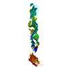
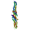
- PDB-1f02: CRYSTAL STRUCTURE OF C-TERMINAL 282-RESIDUE FRAGMENT OF INTIMIN I... -
+ Open data

- Basic information
| Entry | Database: PDB / ID: 1f02 | ||||||
|---|---|---|---|---|---|---|---|
| Title | CRYSTAL STRUCTURE OF C-TERMINAL 282-RESIDUE FRAGMENT OF INTIMIN IN COMPLEX WITH TRANSLOCATED INTIMIN RECEPTOR (TIR) INTIMIN-BINDING DOMAIN | ||||||
 Components Components |
| ||||||
 Keywords Keywords | CELL ADHESION / Immunoglobulin-like fold / C-type lectin-like fold / four-helix bundle | ||||||
| Function / homology |  Function and homology information Function and homology informationpeptidoglycan binding / cell outer membrane / cell adhesion / host cell plasma membrane / protein homodimerization activity / extracellular region / identical protein binding Similarity search - Function | ||||||
| Biological species |  | ||||||
| Method |  X-RAY DIFFRACTION / Resolution: 2.9 Å X-RAY DIFFRACTION / Resolution: 2.9 Å | ||||||
 Authors Authors | Luo, Y. / Frey, E.A. / Pfuetzner, R.A. / Creagh, A.L. / Knoechel, D.G. / Haynes, C.A. / Finlay, B.B. / Strynadka, N.C.J. | ||||||
 Citation Citation | Journal: Nature / Year: 2000 Title: Crystal structure of enteropathogenic Escherichia coli intimin-receptor complex. Authors: Luo, Y. / Frey, E.A. / Pfuetzner, R.A. / Creagh, A.L. / Knoechel, D.G. / Haynes, C.A. / Finlay, B.B. / Strynadka, N.C. | ||||||
| History |
|
- Structure visualization
| Structure viewer | Molecule: MolmilJmol/JSmol |
|---|
- Downloads & links
-Download
| PDBx/mmCIF format | 1f02.cif.gz | 76.5 KB | Display | PDBx/mmCIF format |
|---|---|---|---|---|
| PDB format | pdb1f02.ent.gz | 58 KB | Display | PDB format |
| PDBx/mmJSON format | 1f02.json.gz | Tree view | PDBx/mmJSON format | |
| Others |  Other downloads Other downloads |
-Validation report
| Arichive directory | https://data.pdbj.org/pub/pdb/validation_reports/f0/1f02ftp://data.pdbj.org/pub/pdb/validation_reports/f0/1f02 | HTTPS FTP |
|---|
-Related structure data
| Related structure data |  1f00C  1inm C: citing same article ( |
|---|---|
| Similar structure data |


-Links
 PDBj
PDBj


- Assembly
| Deposited unit | 
| ||||||||
|---|---|---|---|---|---|---|---|---|---|
| 1 | 
| ||||||||
| Unit cell |
|
-Components
| #1: Protein | Mass: 30084.533 Da / Num. of mol.: 1 / Fragment: C-TERMINAL DOMAIN (282 RESIDUES) Source method: isolated from a genetically manipulated source Source: (gene. exp.) |
|---|---|
| #2: Protein | Mass: 7058.585 Da / Num. of mol.: 1 / Fragment: INTIMIN-BINDING DOMAIN Source method: isolated from a genetically manipulated source Source: (gene. exp.) |
| Has protein modification | Y |
-Experimental details
-Experiment
| Experiment | Method: X-RAY DIFFRACTION / Number of used crystals: 2 |
|---|
- Sample preparation
| Crystal | Density Matthews: 5.08 Å3/Da / Density % sol: 75.79 % |
|---|---|
| Crystal grow | Temperature: 295 K / Method: vapor diffusion, hanging drop / pH: 8.5 Details: 1.8 M ammonium sulphate, 200 mM Tris-HCl buffer, pH 8.5, VAPOR DIFFUSION, HANGING DROP, temperature 295.0K |
| Crystal grow | *PLUS Method: vapor diffusion, sitting drop |
| Components of the solutions | *PLUS Conc.: 1.8 M / Details: or 2.5M ammonium sulfate / Chemical formula: K2HPO4 |
-Data collection
| Diffraction | Mean temperature: 100 K |
|---|---|
| Diffraction source | Source: ROTATING ANODE / Type: RIGAKU RU200 / Wavelength: 1.5418 |
| Detector | Type: RIGAKU RAXIS IIC / Detector: IMAGE PLATE / Date: Feb 8, 2000 |
| Radiation | Protocol: SINGLE WAVELENGTH / Monochromatic (M) / Laue (L): M / Scattering type: x-ray |
| Radiation wavelength | Wavelength: 1.5418 Å / Relative weight: 1 |
| Reflection | Resolution: 2.9→20 Å / Num. all: 17457 / Num. obs: 16811 / % possible obs: 96.3 % / Observed criterion σ(F): 0 / Observed criterion σ(I): 0 / Redundancy: 5.08 % / Biso Wilson estimate: 55.9 Å2 / Rmerge(I) obs: 0.095 / Net I/σ(I): 12.2 |
| Reflection shell | Resolution: 2.9→2.96 Å / Redundancy: 2.6 % / Rmerge(I) obs: 0.283 / Num. unique all: 821 / % possible all: 74.9 |
| Reflection shell | *PLUS % possible obs: 74.9 % |
- Processing
| Software |
| |||||||||||||||||||||||||
|---|---|---|---|---|---|---|---|---|---|---|---|---|---|---|---|---|---|---|---|---|---|---|---|---|---|---|
| Refinement | Resolution: 2.9→20 Å / σ(F): 0 / σ(I): 0 / Stereochemistry target values: Engh & Huber
| |||||||||||||||||||||||||
| Refinement step | Cycle: LAST / Resolution: 2.9→20 Å
| |||||||||||||||||||||||||
| Refine LS restraints |
|
