4D HETERONUCLEAR SEPARATED NOE EXPERIMENTS. 3D 13C-SEPARATED/12C-FILTERED NOE EXPERIMENTS FOR REVERSE LABELED AROMATIC SAMPLES
-
Sample preparation
Sample conditions
pH: 7 / Temperature: 313 K
Crystal grow
*PLUS
Method: other / Details: NMR
-
NMR measurement
NMR spectrometer
Type
Manufacturer
Model
Field strength (MHz)
Spectrometer-ID
Bruker AMX500
Bruker
AMX500
500
1
Bruker AMX600
Bruker
AMX600
600
2
-
Processing
Software
Name
Version
Classification
X-PLOR
3.1
modelbuilding
X-PLOR
3.1
refinement
X-PLOR
3.1
phasing
NMR software
Name
Version
Classification
X-PLOR (SEE ABOVE)
ABOVE)
structuresolution
X-PLOR (SEE ABOVE)
ABOVE)
refinement
Refinement
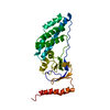
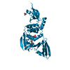
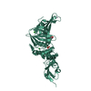
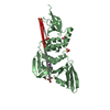
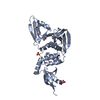
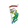
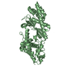
Method: simulated annealing / Software ordinal: 1 Details: THE 3D STRUCTURE OF THE EIN WAS SOLVED BY MULTI-DIMENSIONAL HETERONUCLEAR NMR AND IS BASED ON 4251 EXPERIMENTAL NMR RESTRAINTS: (A) INTRAPROTEIN: 952 SEQUENTIAL (|I- J|=1), 809 MEDIUM RANGE ...Details: THE 3D STRUCTURE OF THE EIN WAS SOLVED BY MULTI-DIMENSIONAL HETERONUCLEAR NMR AND IS BASED ON 4251 EXPERIMENTAL NMR RESTRAINTS: (A) INTRAPROTEIN: 952 SEQUENTIAL (|I- J|=1), 809 MEDIUM RANGE (1 < |I-J| <=5) AND 586 LONG RANGE (|I-J| >5) INTERRESIDUES AND 471 INTRARESIDUE APPROXIMATE INTERPROTON DISTANCE RESTRAINTS; 230 DISTANCES FOR 115 BACKBONE HYDROGEN BONDS; 140 TORSION ANGLE RESTRAINTS; 163 THREE-BOND HN-HA COUPLING CONSTANT RESTRAINTS; AND 498 (258 CALPHA AND 241 CBETA) 13C SHIFT RESTRAINTS. (NUMBERS OF RESIDUES 1 - 259). THE STRUCTURES WERE CALCULATED USING THE SIMULATED ANNEALING PROTOCOL OF NILGES ET AL. (1988) FEBS LETT. 229, 129-136 USING THE PROGRAM X-PLOR 3.1 (BRUNGER) MODIFIED TO INCORPORATE COUPLING CONSTANT (GARRETT ET AL. (1984) J. MAGN. RESON. SERIES B 104, 99-103) AND CARBON CHEMICAL SHIFT (KUSZEWSKI ET AL. (1995) J. MAGN. RESON. SERIES B 106, 92-96) RESTRAINTS, AND A CONFORMATIONAL DATABASE POTENTIAL (KUSZEWSKI ET AL.(1996) PROTEIN SCI. 5, 1067-1080 IN THE RESTRAINED REGULARIZED MEAN COORDINATES THE TEMPERATURE FACTOR FIELD REPRESENTS THE AVERAGE RMS DIFFERENCE BETWEEN THE INDIVIDUAL SIMULATED ANNEALING STRUCTURES AND THE MEAN COORDINATE POSITIONS. THE LAST COLUMN IN THE INDIVIDUAL SA STRUCTURES HAS NO MEANING. BEST FITTING TO GENERATE THE AVERAGE STRUCTURE IS WITH RESPECT TO RESIDUES 1 - 246 (RESIDUES 250 - 259 ARE DISORDERED IN SOLUTION) NOTE THE OCCUPANCY FIELD HAS NO MEANING.
NMR ensemble
Conformers submitted total number: 17
+
About Yorodumi
-
News
-
Feb 9, 2022. New format data for meta-information of EMDB entries
New format data for meta-information of EMDB entries
Version 3 of the EMDB header file is now the official format.
The previous official version 1.9 will be removed from the archive.
In the structure databanks used in Yorodumi, some data are registered as the other names, "COVID-19 virus" and "2019-nCoV". Here are the details of the virus and the list of structure data.
Jan 31, 2019. EMDB accession codes are about to change! (news from PDBe EMDB page)
EMDB accession codes are about to change! (news from PDBe EMDB page)
The allocation of 4 digits for EMDB accession codes will soon come to an end. Whilst these codes will remain in use, new EMDB accession codes will include an additional digit and will expand incrementally as the available range of codes is exhausted. The current 4-digit format prefixed with “EMD-” (i.e. EMD-XXXX) will advance to a 5-digit format (i.e. EMD-XXXXX), and so on. It is currently estimated that the 4-digit codes will be depleted around Spring 2019, at which point the 5-digit format will come into force.
The EM Navigator/Yorodumi systems omit the EMD- prefix.
Related info.:Q: What is EMD? / ID/Accession-code notation in Yorodumi/EM Navigator
Yorodumi is a browser for structure data from EMDB, PDB, SASBDB, etc.
This page is also the successor to EM Navigator detail page, and also detail information page/front-end page for Omokage search.
The word "yorodu" (or yorozu) is an old Japanese word meaning "ten thousand". "mi" (miru) is to see.
Related info.:EMDB / PDB / SASBDB / Comparison of 3 databanks / Yorodumi Search / Aug 31, 2016. New EM Navigator & Yorodumi / Yorodumi Papers / Jmol/JSmol / Function and homology information / Changes in new EM Navigator and Yorodumi
 Movie
Movie Controller

 Yorodumi
 Open data
 Basic information
 Components
 Keywords
 Function and homology information
 Authors
 Citation
 Structure visualization
 Downloads & links
 Other downloads


 PDBj
PDBj
 Assembly
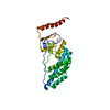
 Sample preparation
 Processing
 X-PLOR