 Movie
Movie Controller


+ Open data

- Basic information
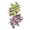
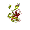
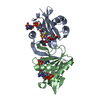
| Entry | Database: PDB / ID: 1exs | ||||||
|---|---|---|---|---|---|---|---|
| Title | STRUCTURE OF PORCINE BETA-LACTOGLOBULIN | ||||||
 Components Components | BETA-LACTOGLOBULIN | ||||||
 Keywords Keywords | LIPID BINDING PROTEIN / lipocalin fold / LIPID-BINDING PROTEIN | ||||||
| Function / homology |  Function and homology information Function and homology information | ||||||
| Biological species |  | ||||||
| Method |  X-RAY DIFFRACTION / Resolution: 2.39 Å X-RAY DIFFRACTION / Resolution: 2.39 Å | ||||||
 Authors Authors | Abrahams, J.P. / Hoedemaeker, F.J. | ||||||
 Citation Citation | Journal: Acta Crystallogr.,Sect.D / Year: 2002 Title: A novel pH-dependent dimerization motif in beta-lactoglobulin from pig (Sus scrofa). Authors: Hoedemaeker, F.J. / Visschers, R.W. / Alting, A.C. / de Kruif, K.G. / Kuil, M.E. / Abrahams, J.P. | ||||||
| History |
|
- Structure visualization
| Structure viewer | Molecule: MolmilJmol/JSmol |
|---|
- Downloads & links
-Download
| PDBx/mmCIF format | 1exs.cif.gz | 48.1 KB | Display | PDBx/mmCIF format |
|---|---|---|---|---|
| PDB format | pdb1exs.ent.gz | 33.5 KB | Display | PDB format |
| PDBx/mmJSON format | 1exs.json.gz | Tree view | PDBx/mmJSON format | |
| Others |  Other downloads Other downloads |
-Validation report
| Arichive directory | https://data.pdbj.org/pub/pdb/validation_reports/ex/1exsftp://data.pdbj.org/pub/pdb/validation_reports/ex/1exs | HTTPS FTP |
|---|
-Related structure data
| Similar structure data |
|---|


-Links
 PDBj
PDBj

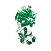
- Assembly
| Deposited unit | 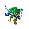
| ||||||||
|---|---|---|---|---|---|---|---|---|---|
| 1 | 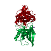
| ||||||||
| Unit cell |
| ||||||||
| Components on special symmetry positions |
|
-Components
| #1: Protein | Mass: 17882.469 Da / Num. of mol.: 1 / Fragment: VARIANT 1A / Source method: isolated from a natural source Details: A PUTATIVE BUT BIOLOGICALLY FUNCTIONAL DIMER IS CREATED BY THE CRYSTALLOGRAPHIC 2-FOLD AXIS Source: (natural) |
|---|---|
| #2: Chemical | ChemComp-NA /   Mass: 22.990 Da / Num. of mol.: 1 / Source method: obtained synthetically / Formula: Na Mass: 22.990 Da / Num. of mol.: 1 / Source method: obtained synthetically / Formula: Na |
| #3: Chemical | ChemComp-GOL /   Mass: 92.094 Da / Num. of mol.: 1 / Source method: obtained synthetically / Formula: C3H8O3 Mass: 92.094 Da / Num. of mol.: 1 / Source method: obtained synthetically / Formula: C3H8O3 |
| #4: Water | ChemComp-HOH /  Mass: 18.015 Da / Num. of mol.: 154 / Source method: isolated from a natural source / Formula: H2O Mass: 18.015 Da / Num. of mol.: 154 / Source method: isolated from a natural source / Formula: H2O |
| Has protein modification | Y |
-Experimental details
-Experiment
| Experiment | Method: X-RAY DIFFRACTION / Number of used crystals: 2 |
|---|
- Sample preparation
| Crystal | Density Matthews: 4.12 Å3/Da / Density % sol: 70.12 % | ||||||||||||||||||||||||||||
|---|---|---|---|---|---|---|---|---|---|---|---|---|---|---|---|---|---|---|---|---|---|---|---|---|---|---|---|---|---|
| Crystal grow | Temperature: 293 K / Method: microbatch / pH: 3.2 Details: Sodium chloride, Formate buffer, pH 3.2, microbatch, temperature 293K | ||||||||||||||||||||||||||||
| Crystal grow | *PLUS Method: batch method | ||||||||||||||||||||||||||||
| Components of the solutions | *PLUS
|
-Data collection
| Diffraction | Mean temperature: 100 K |
|---|---|
| Diffraction source | Source: ROTATING ANODE / Type: ENRAF-NONIUS FR591 / Wavelength: 1.5418 |
| Detector | Type: MARRESEARCH / Detector: IMAGE PLATE / Date: Mar 15, 2000 |
| Radiation | Protocol: SINGLE WAVELENGTH / Monochromatic (M) / Laue (L): M / Scattering type: x-ray |
| Radiation wavelength | Wavelength: 1.5418 Å / Relative weight: 1 |
| Reflection | Resolution: 2.38→999 Å / Num. all: 12250 / Num. obs: 12225 / % possible obs: 99.8 % / Observed criterion σ(F): 0 / Observed criterion σ(I): 2.7 / Redundancy: 10.2 % / Biso Wilson estimate: 55 Å2 / Rmerge(I) obs: 0.052 / Net I/σ(I): 9.7 |
| Reflection shell | Resolution: 2.38→2.44 Å / Redundancy: 9.4 % / Rmerge(I) obs: 0.293 / Num. unique all: 870 / % possible all: 97.3 |
| Reflection | *PLUS Highest resolution: 2.39 Å / Lowest resolution: 100 Å / Num. measured all: 124487 |
| Reflection shell | *PLUS Highest resolution: 2.39 Å / % possible obs: 97.3 % / Num. unique obs: 870 / Num. measured obs: 8192 / Rmerge(I) obs: 0.25 / Mean I/σ(I) obs: 2.7 |
- Processing
| Software |
| ||||||||||||||||||||||||
|---|---|---|---|---|---|---|---|---|---|---|---|---|---|---|---|---|---|---|---|---|---|---|---|---|---|
| Refinement | Resolution: 2.39→40 Å / Cross valid method: THROUGHOUT / σ(F): 0 / σ(I): 0 / Stereochemistry target values: Engh & Huber / Details: MLKF CDIR
| ||||||||||||||||||||||||
| Refinement step | Cycle: LAST / Resolution: 2.39→40 Å
| ||||||||||||||||||||||||
| Refine LS restraints |
| ||||||||||||||||||||||||
| Software | *PLUS Name: REFMAC/ARP_WARP / Classification: refinement | ||||||||||||||||||||||||
| Refinement | *PLUS σ(F): 0 / Rfactor obs: 0.218 | ||||||||||||||||||||||||
| Solvent computation | *PLUS | ||||||||||||||||||||||||
| Displacement parameters | *PLUS | ||||||||||||||||||||||||
| Refine LS restraints | *PLUS
| ||||||||||||||||||||||||
| LS refinement shell | *PLUS Highest resolution: 2.38 Å / Lowest resolution: 2.51 Å / Rfactor Rfree: 0.318 / Num. reflection Rfree: 83 / Rfactor Rwork: 0.258 / Num. reflection Rwork: 1497 |
