 Movie
Movie Controller


+ Open data

- Basic information
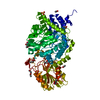
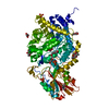
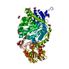
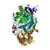
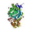
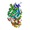
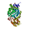
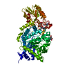
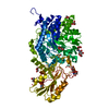
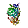
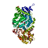
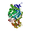
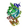
| Entry | Database: PDB / ID: 1ex1 | |||||||||
|---|---|---|---|---|---|---|---|---|---|---|
| Title | BETA-D-GLUCAN EXOHYDROLASE FROM BARLEY | |||||||||
 Components Components | PROTEIN (BETA-D-GLUCAN EXOHYDROLASE ISOENZYME EXO1) | |||||||||
 Keywords Keywords | HYDROLASE / CELL WALL DEGRADATION | |||||||||
| Function / homology |  Function and homology information Function and homology informationhydrolase activity, hydrolyzing O-glycosyl compounds / carbohydrate metabolic process Similarity search - Function | |||||||||
| Biological species |  | |||||||||
| Method |  X-RAY DIFFRACTION / MIRAS / Resolution: 2.2 Å X-RAY DIFFRACTION / MIRAS / Resolution: 2.2 Å | |||||||||
 Authors Authors | Varghese, J.N. / Hrmova, M. / Fincher, G.B. | |||||||||
 Citation Citation | Journal: Structure Fold.Des. / Year: 1999 Title: Three-dimensional structure of a barley beta-D-glucan exohydrolase, a family 3 glycosyl hydrolase. Authors: Varghese, J.N. / Hrmova, M. / Fincher, G.B. #1: Journal: Acta Crystallogr.,Sect.D / Year: 1998Title: Crystallization and Preliminary X-Ray Analysis of Beta-D-Glucan Exohydrolase Isoenzyme Exo1 from Barley (Hordeum Vulgare); a Family 3 Glycosyl Hydrolase Authors: Varghese, J.N. / Hrmova, M. / Hoj, P.B. / Fincher, G.B. | |||||||||
| History |
|
- Structure visualization
| Structure viewer | Molecule: MolmilJmol/JSmol |
|---|
- Downloads & links
-Download
| PDBx/mmCIF format | 1ex1.cif.gz | 133 KB | Display | PDBx/mmCIF format |
|---|---|---|---|---|
| PDB format | pdb1ex1.ent.gz | 102.2 KB | Display | PDB format |
| PDBx/mmJSON format | 1ex1.json.gz | Tree view | PDBx/mmJSON format | |
| Others |  Other downloads Other downloads |
-Validation report
| Summary document | 1ex1_validation.pdf.gz | 901.6 KB | Display | wwPDB validaton report |
|---|---|---|---|---|
| Full document | 1ex1_full_validation.pdf.gz | 916.1 KB | Display | |
| Data in XML | 1ex1_validation.xml.gz | 27 KB | Display | |
| Data in CIF | 1ex1_validation.cif.gz | 37.8 KB | Display | |
| Arichive directory | https://data.pdbj.org/pub/pdb/validation_reports/ex/1ex1ftp://data.pdbj.org/pub/pdb/validation_reports/ex/1ex1 | HTTPS FTP |
-Related structure data
| Similar structure data |
|---|


-Links
 PDBj
PDBj

- Assembly
| Deposited unit | 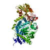
| ||||||||
|---|---|---|---|---|---|---|---|---|---|
| 1 |
| ||||||||
| Unit cell |
|
-Components
| #1: Protein | Mass: 65475.617 Da / Num. of mol.: 1 / Source method: isolated from a natural source Details: THREE GLYCSYLATED SITES AT ASN 221, 498, 600 GLUCOSE BOUND IN PUTATIVE ACTIVE SITE Source: (natural) References: GenBank: AAD23382, UniProt: Q9XEI3*PLUS, glucan 1,3-beta-glucosidase |
|---|---|
| #2: Polysaccharide | 2-acetamido-2-deoxy-beta-D-glucopyranose-(1-2)-alpha-D-mannopyranose-(1-6)-alpha-D-mannopyranose-(1- ...2-acetamido-2-deoxy-beta-D-glucopyranose-(1-2)-alpha-D-mannopyranose-(1-6)-alpha-D-mannopyranose-(1-4)-2-acetamido-2-deoxy-beta-D-glucopyranose-(1-4)-[alpha-D-fucopyranose-(1-3)]2-acetamido-2-deoxy-beta-D-glucopyranose Source method: isolated from a genetically manipulated source |
| #3: Sugar | ChemComp-NAG /   Type: D-saccharide, beta linking / Mass: 221.208 Da / Num. of mol.: 1 Type: D-saccharide, beta linking / Mass: 221.208 Da / Num. of mol.: 1Source method: isolated from a genetically manipulated source Formula: C8H15NO6 |
| #4: Sugar | ChemComp-GLC /   Type: D-saccharide, alpha linking / Mass: 180.156 Da / Num. of mol.: 1 Type: D-saccharide, alpha linking / Mass: 180.156 Da / Num. of mol.: 1Source method: isolated from a genetically manipulated source Formula: C6H12O6 |
| #5: Water | ChemComp-HOH /  Mass: 18.015 Da / Num. of mol.: 220 / Source method: isolated from a natural source / Formula: H2O Mass: 18.015 Da / Num. of mol.: 220 / Source method: isolated from a natural source / Formula: H2O |
| Has protein modification | Y |
-Experimental details
-Experiment
| Experiment | Method: X-RAY DIFFRACTION / Number of used crystals: 1 |
|---|
- Sample preparation
| Crystal | Density Matthews: 1.83 Å3/Da / Density % sol: 66 % Description: THE SAME CRYSTAL WAS USED TO COLLECT BOTH NATIVE AND PIP DATA | ||||||||||||||||||||||||||||||||||||||||
|---|---|---|---|---|---|---|---|---|---|---|---|---|---|---|---|---|---|---|---|---|---|---|---|---|---|---|---|---|---|---|---|---|---|---|---|---|---|---|---|---|---|
| Crystal grow | pH: 7 / Details: pH 7.0 | ||||||||||||||||||||||||||||||||||||||||
| Crystal | *PLUS | ||||||||||||||||||||||||||||||||||||||||
| Crystal grow | *PLUS Temperature: 277-279 K / Method: vapor diffusion, hanging drop / Details: Hrmova, M., (1998) Acta Cryst., D54, 687. | ||||||||||||||||||||||||||||||||||||||||
| Components of the solutions | *PLUS
|
-Data collection
| Diffraction | Mean temperature: 291 K |
|---|---|
| Diffraction source | Source: ROTATING ANODE / Type: MACSCIENCE M18X / Wavelength: 1.5418 |
| Detector | Type: RIGAKU RAXIS II / Detector: IMAGE PLATE / Date: May 15, 1997 / Details: MIRRORS |
| Radiation | Monochromator: NI FILTER / Protocol: SINGLE WAVELENGTH / Monochromatic (M) / Laue (L): M / Scattering type: x-ray |
| Radiation wavelength | Wavelength: 1.5418 Å / Relative weight: 1 |
| Reflection | Resolution: 2.2→20 Å / Num. obs: 408526 / % possible obs: 78 % / Observed criterion σ(I): 1 / Redundancy: 2.5 % / Biso Wilson estimate: 26.6 Å2 / Rmerge(I) obs: 0.054 / Net I/σ(I): 20.1 |
| Reflection shell | Resolution: 2.2→2.3 Å / Rmerge(I) obs: 0.142 / Mean I/σ(I) obs: 2.8 / % possible all: 37.8 |
| Reflection | *PLUS Num. obs: 40310 / Observed criterion σ(I): 1 / Redundancy: 3.7 % / Num. measured all: 148526 |
| Reflection shell | *PLUS % possible obs: 32 % |
- Processing
| Software |
| ||||||||||||||||||||||||||||||||||||||||||||||||||||||||||||||||||||||||||||||||||||
|---|---|---|---|---|---|---|---|---|---|---|---|---|---|---|---|---|---|---|---|---|---|---|---|---|---|---|---|---|---|---|---|---|---|---|---|---|---|---|---|---|---|---|---|---|---|---|---|---|---|---|---|---|---|---|---|---|---|---|---|---|---|---|---|---|---|---|---|---|---|---|---|---|---|---|---|---|---|---|---|---|---|---|---|---|---|
| Refinement | Method to determine structure: MIRAS / Resolution: 2.2→20 Å / SU B: 4.29 / Cross valid method: THROUGHOUT / σ(F): 1
| ||||||||||||||||||||||||||||||||||||||||||||||||||||||||||||||||||||||||||||||||||||
| Refinement step | Cycle: LAST / Resolution: 2.2→20 Å
| ||||||||||||||||||||||||||||||||||||||||||||||||||||||||||||||||||||||||||||||||||||
| Refine LS restraints |
| ||||||||||||||||||||||||||||||||||||||||||||||||||||||||||||||||||||||||||||||||||||
| Software | *PLUS Name: REFMAC / Classification: refinement | ||||||||||||||||||||||||||||||||||||||||||||||||||||||||||||||||||||||||||||||||||||
| Refinement | *PLUS Highest resolution: 2.2 Å / σ(F): 1 / % reflection Rfree: 5 % / Rfactor obs: 0.17 / Rfactor Rwork: 0.17 | ||||||||||||||||||||||||||||||||||||||||||||||||||||||||||||||||||||||||||||||||||||
| Solvent computation | *PLUS | ||||||||||||||||||||||||||||||||||||||||||||||||||||||||||||||||||||||||||||||||||||
| Displacement parameters | *PLUS | ||||||||||||||||||||||||||||||||||||||||||||||||||||||||||||||||||||||||||||||||||||
| Refine LS restraints | *PLUS
| ||||||||||||||||||||||||||||||||||||||||||||||||||||||||||||||||||||||||||||||||||||
| LS refinement shell | *PLUS Rfactor Rfree: 0.283 / Rfactor Rwork: 0.202 / Num. reflection obs: 1633 / Rfactor obs: 0.202 |
