 Movie
Movie Controller

[English] 日本語
 Yorodumi
- PDB-1iex: Crystal structure of barley beta-D-glucan glucohydrolase isoenzym... -
+ Open data

- Basic information
| Entry | Database: PDB / ID: 1iex | |||||||||
|---|---|---|---|---|---|---|---|---|---|---|
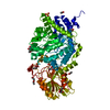
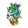
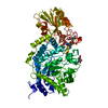
| Title | Crystal structure of barley beta-D-glucan glucohydrolase isoenzyme Exo1 in complex with 4I,4III,4V-S-trithiocellohexaose | |||||||||
 Components Components | BETA-D-GLUCAN GLUCOHYDROLASE ISOENZYME EXO1 | |||||||||
 Keywords Keywords | HYDROLASE / 2-domain fold | |||||||||
| Function / homology |  Function and homology information Function and homology informationhydrolase activity, hydrolyzing O-glycosyl compounds / carbohydrate metabolic process Similarity search - Function | |||||||||
| Biological species |  | |||||||||
| Method |  X-RAY DIFFRACTION / SYNCHROTRON / MOLECULAR REPLACEMENT / Resolution: 2.2 Å X-RAY DIFFRACTION / SYNCHROTRON / MOLECULAR REPLACEMENT / Resolution: 2.2 Å | |||||||||
 Authors Authors | Hrmova, M. / DeGori, R. / Fincher, G.B. / Smith, B.J. / Driguez, H. / Varghese, J.N. | |||||||||
 Citation Citation | Journal: Structure / Year: 2001 Title: Catalytic mechanisms and reaction intermediates along the hydrolytic pathway of a plant beta-D-glucan glucohydrolase. Authors: Hrmova, M. / Varghese, J.N. / De Gori, R. / Smith, B.J. / Driguez, H. / Fincher, G.B. | |||||||||
| History |
| |||||||||
| Remark 600 | HETEROGEN The ligand 4I,4III,4V-S-trithiocellohexaose is present in the crystal. Only the ...HETEROGEN The ligand 4I,4III,4V-S-trithiocellohexaose is present in the crystal. Only the thiocellobiose portion of the ligand is seen in the density; the 4III,4V-S-thiocellotetraose moiety is disordered and can not be seen. | |||||||||
| Remark 999 | SEQUENCE The sequence in the GenBank entry might be a sequencing error at residue 345. The electron ...SEQUENCE The sequence in the GenBank entry might be a sequencing error at residue 345. The electron density unambiguously proved the presence of LYS instead of ASN at this residue. |
- Structure visualization
| Structure viewer | Molecule: MolmilJmol/JSmol |
|---|
- Downloads & links
-Download
| PDBx/mmCIF format | 1iex.cif.gz | 136.5 KB | Display | PDBx/mmCIF format |
|---|---|---|---|---|
| PDB format | pdb1iex.ent.gz | 104.1 KB | Display | PDB format |
| PDBx/mmJSON format | 1iex.json.gz | Tree view | PDBx/mmJSON format | |
| Others |  Other downloads Other downloads |
-Validation report
| Arichive directory | https://data.pdbj.org/pub/pdb/validation_reports/ie/1iexftp://data.pdbj.org/pub/pdb/validation_reports/ie/1iex | HTTPS FTP |
|---|
-Related structure data
| Related structure data | 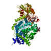 1ieqC  1ievC  1iewC  1ex1S S: Starting model for refinement C: citing same article ( |
|---|---|
| Similar structure data |

-Links
 PDBj
PDBj- Assembly
| Deposited unit | 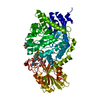
| ||||||||
|---|---|---|---|---|---|---|---|---|---|
| 1 |
| ||||||||
| Unit cell |
| ||||||||
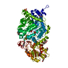
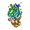
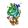
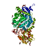
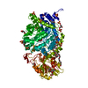
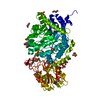
| Details | The biological assembly is a monomer constructed from an (alpha/beta)8 barrel and an (alpha/beta)6 sandwich. |
-Components
| #1: Protein | Mass: 65475.617 Da / Num. of mol.: 1 / Source method: isolated from a natural source / Source: (natural) References: GenBank: 4566505, UniProt: Q9XEI3*PLUS, glucan 1,3-beta-glucosidase |
|---|---|
| #2: Polysaccharide | 2-acetamido-2-deoxy-beta-D-glucopyranose-(1-2)-alpha-D-mannopyranose-(1-6)-beta-D-mannopyranose-(1- ...2-acetamido-2-deoxy-beta-D-glucopyranose-(1-2)-alpha-D-mannopyranose-(1-6)-beta-D-mannopyranose-(1-4)-2-acetamido-2-deoxy-beta-D-glucopyranose-(1-4)-[alpha-L-fucopyranose-(1-3)]2-acetamido-2-deoxy-beta-D-glucopyranose Source method: isolated from a genetically manipulated source |
| #3: Polysaccharide | beta-D-glucopyranose-(1-4)-4-thio-beta-D-glucopyranose / thio-beta-cellobiose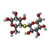  Type: oligosaccharide, Oligosaccharide / Class: Substrate analog / Mass: 358.362 Da / Num. of mol.: 1 Type: oligosaccharide, Oligosaccharide / Class: Substrate analog / Mass: 358.362 Da / Num. of mol.: 1Source method: isolated from a genetically manipulated source Details: oligosaccharide with S-glycosidic bond between monosaccharides References: thio-beta-cellobiose |
| #4: Sugar | ChemComp-NAG /   Type: D-saccharide, beta linking / Mass: 221.208 Da / Num. of mol.: 1 Type: D-saccharide, beta linking / Mass: 221.208 Da / Num. of mol.: 1Source method: isolated from a genetically manipulated source Formula: C8H15NO6 |
| #5: Water | ChemComp-HOH /  Mass: 18.015 Da / Num. of mol.: 301 / Source method: isolated from a natural source / Formula: H2O Mass: 18.015 Da / Num. of mol.: 301 / Source method: isolated from a natural source / Formula: H2O |
| Has protein modification | Y |
-Experimental details
-Experiment
| Experiment | Method: X-RAY DIFFRACTION / Number of used crystals: 3 |
|---|
- Sample preparation
| Crystal | Density Matthews: 3.71 Å3/Da / Density % sol: 66.87 % | ||||||||||||||||||||||||||||||||||||||||
|---|---|---|---|---|---|---|---|---|---|---|---|---|---|---|---|---|---|---|---|---|---|---|---|---|---|---|---|---|---|---|---|---|---|---|---|---|---|---|---|---|---|
| Crystal grow | Temperature: 277 K / Method: vapor diffusion, hanging drop / pH: 7 Details: ammonium sulfate, PEG 400, sodium acetate, Hepes-NaOH, pH 7.0, VAPOR DIFFUSION, HANGING DROP, temperature 277K | ||||||||||||||||||||||||||||||||||||||||
| Crystal grow | *PLUS Temperature: 277-279 K / Details: Hrmova, M., (1998) Acta Cryst., D54, 687. | ||||||||||||||||||||||||||||||||||||||||
| Components of the solutions | *PLUS
|
-Data collection
| Diffraction | Mean temperature: 255 K |
|---|---|
| Diffraction source | Source: SYNCHROTRON / Site: Photon Factory  / Beamline: BL-6A / Wavelength: 1 Å / Beamline: BL-6A / Wavelength: 1 Å |
| Detector | Type: WEISSENBERG / Detector: DIFFRACTOMETER / Date: Mar 1, 1998 / Details: Collimator |
| Radiation | Protocol: SINGLE WAVELENGTH / Monochromatic (M) / Laue (L): M / Scattering type: x-ray |
| Radiation wavelength | Wavelength: 1 Å / Relative weight: 1 |
| Reflection | Resolution: 2.2→20 Å / Num. all: 123698 / Num. obs: 46444 / % possible obs: 83.4 % / Observed criterion σ(F): 0 / Observed criterion σ(I): 0 / Redundancy: 4.4 % / Rmerge(I) obs: 0.098 / Net I/σ(I): 15.8 |
| Reflection shell | Resolution: 2.2→20 Å / Rmerge(I) obs: 0.486 / % possible all: 55.8 |
| Reflection | *PLUS Redundancy: 4.4 % / Num. measured all: 123698 |
- Processing
| Software |
| ||||||||||||||||||||
|---|---|---|---|---|---|---|---|---|---|---|---|---|---|---|---|---|---|---|---|---|---|
| Refinement | Method to determine structure: MOLECULAR REPLACEMENT Starting model: PDB ENTRY 1EX1 Resolution: 2.2→12 Å / Isotropic thermal model: Isotropic / Cross valid method: THROUGHOUT / σ(F): 0 / σ(I): 0 / Stereochemistry target values: Engh & Huber
| ||||||||||||||||||||
| Refine analyze | Luzzati coordinate error obs: 0.24 Å | ||||||||||||||||||||
| Refinement step | Cycle: LAST / Resolution: 2.2→12 Å
| ||||||||||||||||||||
| Refine LS restraints |
| ||||||||||||||||||||
| Software | *PLUS Name: CNS / Classification: refinement | ||||||||||||||||||||
| Refinement | *PLUS Highest resolution: 2.2 Å / Lowest resolution: 12 Å / σ(F): 0 / Rfactor obs: 0.1776 / Rfactor Rfree: 0.2109 | ||||||||||||||||||||
| Solvent computation | *PLUS | ||||||||||||||||||||
| Displacement parameters | *PLUS | ||||||||||||||||||||
| Refine LS restraints | *PLUS
|
