 Movie
Movie Controller

[English] 日本語
 Yorodumi
- PDB-1euj: A NOVEL ANTI-TUMOR CYTOKINE CONTAINS A RNA-BINDING MOTIF PRESENT ... -
+ Open data

- Basic information
| Entry | Database: PDB / ID: 1euj | ||||||
|---|---|---|---|---|---|---|---|
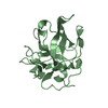
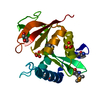
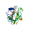
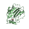
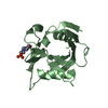
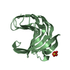
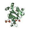
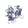
| Title | A NOVEL ANTI-TUMOR CYTOKINE CONTAINS A RNA-BINDING MOTIF PRESENT IN AMINOACYL-TRNA SYNTHETASES | ||||||
 Components Components | ENDOTHELIAL MONOCYTE ACTIVATING POLYPEPTIDE 2 | ||||||
 Keywords Keywords | CYTOKINE / EMAP 2 / EMAP II / tRNA synthetase / apoptosis / RNA binding motif | ||||||
| Function / homology |  Function and homology information Function and homology informationpositive regulation of glucagon secretion / Selenoamino acid metabolism / Cytosolic tRNA aminoacylation / aminoacyl-tRNA synthetase multienzyme complex / leukocyte migration / negative regulation of endothelial cell proliferation / Transcriptional and post-translational regulation of MITF-M expression and activity / cytokine activity / cell-cell signaling / GTPase binding ...positive regulation of glucagon secretion / Selenoamino acid metabolism / Cytosolic tRNA aminoacylation / aminoacyl-tRNA synthetase multienzyme complex / leukocyte migration / negative regulation of endothelial cell proliferation / Transcriptional and post-translational regulation of MITF-M expression and activity / cytokine activity / cell-cell signaling / GTPase binding / angiogenesis / defense response to virus / tRNA binding / translation / inflammatory response / apoptotic process / cell surface / endoplasmic reticulum / Golgi apparatus / protein homodimerization activity / : / membrane / nucleus / cytosol Similarity search - Function | ||||||
| Biological species |  Homo sapiens (human) Homo sapiens (human) | ||||||
| Method |  X-RAY DIFFRACTION / Resolution: 1.8 Å X-RAY DIFFRACTION / Resolution: 1.8 Å | ||||||
 Authors Authors | Kim, Y. / Shin, J. / Li, R. / Cheong, C. / Kim, S. | ||||||
 Citation Citation | Journal: J.Biol.Chem. / Year: 2000 Title: A novel anti-tumor cytokine contains an RNA binding motif present in aminoacyl-tRNA synthetases. Authors: Kim, Y. / Shin, J. / Li, R. / Cheong, C. / Kim, K. / Kim, S. | ||||||
| History |
|
- Structure visualization
| Structure viewer | Molecule: MolmilJmol/JSmol |
|---|
- Downloads & links
-Download
| PDBx/mmCIF format | 1euj.cif.gz | 75.7 KB | Display | PDBx/mmCIF format |
|---|---|---|---|---|
| PDB format | pdb1euj.ent.gz | 57.2 KB | Display | PDB format |
| PDBx/mmJSON format | 1euj.json.gz | Tree view | PDBx/mmJSON format | |
| Others |  Other downloads Other downloads |
-Validation report
| Arichive directory | https://data.pdbj.org/pub/pdb/validation_reports/eu/1eujftp://data.pdbj.org/pub/pdb/validation_reports/eu/1euj | HTTPS FTP |
|---|
-Related structure data
| Similar structure data |
|---|


-Links
 PDBj
PDBj
- Assembly
| Deposited unit | 
| ||||||||
|---|---|---|---|---|---|---|---|---|---|
| 1 | 
| ||||||||
| 2 | 
| ||||||||
| Unit cell |
| ||||||||
| Details | The biological assembly is a monomer in vivo, but forms a dimer in a asymmetric unit in crystal by the two-fold. |
-Components
| #1: Protein | Mass: 18242.178 Da / Num. of mol.: 2 Source method: isolated from a genetically manipulated source Source: (gene. exp.) Homo sapiens (human) / Production host:  #2: Water | ChemComp-HOH / |  Mass: 18.015 Da / Num. of mol.: 193 / Source method: isolated from a natural source / Formula: H2O Mass: 18.015 Da / Num. of mol.: 193 / Source method: isolated from a natural source / Formula: H2O |
|---|
-Experimental details
-Experiment
| Experiment | Method: X-RAY DIFFRACTION / Number of used crystals: 1 |
|---|
- Sample preparation
| Crystal | Density Matthews: 2.62 Å3/Da / Density % sol: 53.14 % | ||||||||||||||||||||||||||||||||||||||||||||||||||||||
|---|---|---|---|---|---|---|---|---|---|---|---|---|---|---|---|---|---|---|---|---|---|---|---|---|---|---|---|---|---|---|---|---|---|---|---|---|---|---|---|---|---|---|---|---|---|---|---|---|---|---|---|---|---|---|---|
| Crystal grow | Temperature: 294 K / Method: vapor diffusion, hanging drop / pH: 4.6 Details: 20% PEG 4000, 100 mM NaAcetate, 15 mM MgCl2, pH 4.6, VAPOR DIFFUSION, HANGING DROP, temperature 21K | ||||||||||||||||||||||||||||||||||||||||||||||||||||||
| Crystal | *PLUS Density % sol: 64 % | ||||||||||||||||||||||||||||||||||||||||||||||||||||||
| Crystal grow | *PLUS pH: 8 | ||||||||||||||||||||||||||||||||||||||||||||||||||||||
| Components of the solutions | *PLUS
|
-Data collection
| Diffraction | Mean temperature: 298 K |
|---|---|
| Diffraction source | Source: ROTATING ANODE / Type: RIGAKU RU200 / Wavelength: 1.5418 |
| Detector | Type: RIGAKU RAXIS IV / Detector: IMAGE PLATE / Date: Nov 13, 1998 |
| Radiation | Protocol: SINGLE WAVELENGTH / Monochromatic (M) / Laue (L): M / Scattering type: x-ray |
| Radiation wavelength | Wavelength: 1.5418 Å / Relative weight: 1 |
| Reflection | Resolution: 1.8→20 Å / Num. all: 35728 / Num. obs: 31502 / % possible obs: 88.2 % / Observed criterion σ(F): 0 / Observed criterion σ(I): 1.8 / Redundancy: 3.1 % / Biso Wilson estimate: 15 Å2 / Rmerge(I) obs: 0.068 / Net I/σ(I): 31.3 |
| Reflection shell | Resolution: 1.8→1.89 Å / Redundancy: 1.4 % / Rmerge(I) obs: 0.282 / % possible all: 37.9 |
| Reflection | *PLUS Num. measured all: 96335 |
| Reflection shell | *PLUS % possible obs: 37.9 % / Mean I/σ(I) obs: 1.8 |
- Processing
| Software |
| ||||||||||||||||||||
|---|---|---|---|---|---|---|---|---|---|---|---|---|---|---|---|---|---|---|---|---|---|
| Refinement | Resolution: 1.8→20 Å / σ(F): 0 / σ(I): 0 / Stereochemistry target values: Engh & Huber
| ||||||||||||||||||||
| Refinement step | Cycle: LAST / Resolution: 1.8→20 Å
| ||||||||||||||||||||
| Refine LS restraints |
| ||||||||||||||||||||
| Software | *PLUS Name: CNS / Classification: refinement | ||||||||||||||||||||
| Refinement | *PLUS Highest resolution: 1.8 Å / Lowest resolution: 20 Å / σ(F): 0 / Rfactor obs: 0.208 | ||||||||||||||||||||
| Solvent computation | *PLUS | ||||||||||||||||||||
| Displacement parameters | *PLUS Biso mean: 31.7 Å2 |
