 Movie
Movie Controller

[English] 日本語
 Yorodumi
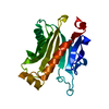
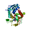
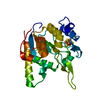
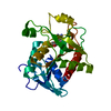
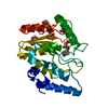
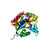
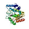
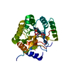
- PDB-1eq6: 1.9 ANGSTROM RESOLUTION CRYSTAL STRUCTURE OF THE SACCHAROMYCES CE... -
+ Open data

- Basic information
| Entry | Database: PDB / ID: 1eq6 | ||||||
|---|---|---|---|---|---|---|---|
| Title | 1.9 ANGSTROM RESOLUTION CRYSTAL STRUCTURE OF THE SACCHAROMYCES CEREVISIAE RAN-BINDING PROTEIN MOG1P | ||||||
 Components Components | MOG1P | ||||||
 Keywords Keywords | PROTEIN TRANSPORT / alpha-beta | ||||||
| Function / homology |  Function and homology information Function and homology informationmRNA transport / guanyl-nucleotide exchange factor activity / small GTPase binding / protein import into nucleus / nucleus Similarity search - Function | ||||||
| Biological species |  | ||||||
| Method |  X-RAY DIFFRACTION / SYNCHROTRON / MAD PLUS SIR / Resolution: 1.9 Å X-RAY DIFFRACTION / SYNCHROTRON / MAD PLUS SIR / Resolution: 1.9 Å | ||||||
 Authors Authors | Stewart, M. / Baker, R.P. | ||||||
 Citation Citation | Journal: J.Mol.Biol. / Year: 2000 Title: 1.9 A resolution crystal structure of the Saccharomyces cerevisiae Ran-binding protein Mog1p. Authors: Stewart, M. / Baker, R.P. | ||||||
| History |
|
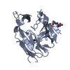
- Structure visualization
| Structure viewer | Molecule: MolmilJmol/JSmol |
|---|
- Downloads & links
-Download
| PDBx/mmCIF format | 1eq6.cif.gz | 51.4 KB | Display | PDBx/mmCIF format |
|---|---|---|---|---|
| PDB format | pdb1eq6.ent.gz | 36.6 KB | Display | PDB format |
| PDBx/mmJSON format | 1eq6.json.gz | Tree view | PDBx/mmJSON format | |
| Others |  Other downloads Other downloads |
-Validation report
| Arichive directory | https://data.pdbj.org/pub/pdb/validation_reports/eq/1eq6ftp://data.pdbj.org/pub/pdb/validation_reports/eq/1eq6 | HTTPS FTP |
|---|
-Related structure data
| Similar structure data |
|---|
-Links
 PDBj
PDBj
- Assembly
| Deposited unit | 
| ||||||||
|---|---|---|---|---|---|---|---|---|---|
| 1 |
| ||||||||
| Unit cell |
|
-Components
| #1: Protein | Mass: 21160.680 Da / Num. of mol.: 1 / Fragment: RESIDUES 30-218 Source method: isolated from a genetically manipulated source Source: (gene. exp.) Plasmid: PMW172 / Production host:  |
|---|---|
| #2: Water | ChemComp-HOH /  Mass: 18.015 Da / Num. of mol.: 124 / Source method: isolated from a natural source / Formula: H2O Mass: 18.015 Da / Num. of mol.: 124 / Source method: isolated from a natural source / Formula: H2O |
-Experimental details
-Experiment
| Experiment | Method: X-RAY DIFFRACTION / Number of used crystals: 1 |
|---|
- Sample preparation
| Crystal | Density Matthews: 2.73 Å3/Da / Density % sol: 48.5 % / Description: 3A PHASES OBTAINED FROM SE-MET MAD PLUS Hg SIR | |||||||||||||||||||||||||
|---|---|---|---|---|---|---|---|---|---|---|---|---|---|---|---|---|---|---|---|---|---|---|---|---|---|---|
| Crystal grow | Temperature: 291 K / Method: vapor diffusion, hanging drop / pH: 6 Details: PEG4000, glycerol, sodium acetate, DTT, pH 6.0, VAPOR DIFFUSION, HANGING DROP, temperature 18K | |||||||||||||||||||||||||
| Crystal | *PLUS Density % sol: 49.5 % | |||||||||||||||||||||||||
| Crystal grow | *PLUS Temperature: 291 K / pH: 5.6 / Method: vapor diffusionDetails: Baker, R.P.,(2000) Acta Crystallog. Sect., D56, 229. | |||||||||||||||||||||||||
| Components of the solutions | *PLUS
|
-Data collection
| Diffraction | Mean temperature: 100 K |
|---|---|
| Diffraction source | Source: SYNCHROTRON / Site: ESRF  / Beamline: ID2 / Wavelength: 0.9887 / Beamline: ID2 / Wavelength: 0.9887 |
| Detector | Type: MARRESEARCH / Detector: IMAGE PLATE / Date: May 15, 1999 |
| Radiation | Protocol: SINGLE WAVELENGTH / Monochromatic (M) / Laue (L): M / Scattering type: x-ray |
| Radiation wavelength | Wavelength: 0.9887 Å / Relative weight: 1 |
| Reflection | Resolution: 1.9→20 Å / Num. all: 18951 / Num. obs: 18625 / % possible obs: 98.3 % / Observed criterion σ(F): 0 / Observed criterion σ(I): 0 / Redundancy: 3.5 % / Biso Wilson estimate: 33 Å2 / Rmerge(I) obs: 0.039 / Net I/σ(I): 11.5 |
| Reflection shell | Resolution: 1.9→2 Å / Redundancy: 3.3 % / Mean I/σ(I) obs: 4.9 / Num. unique all: 2679 / Rsym value: 0.146 / % possible all: 98.5 |
| Reflection shell | *PLUS % possible obs: 98.7 % / Rmerge(I) obs: 0.146 |
- Processing
| Software |
| |||||||||||||||||||||||||||
|---|---|---|---|---|---|---|---|---|---|---|---|---|---|---|---|---|---|---|---|---|---|---|---|---|---|---|---|---|
| Refinement | Method to determine structure: MAD PLUS SIR / Resolution: 1.9→20 Å / Cross valid method: FREE-R / σ(F): 0 / σ(I): 0 / Stereochemistry target values: ENGH & HUBER / Details: used maximum likelihood target "mlf"
| |||||||||||||||||||||||||||
| Displacement parameters | Biso mean: 33.32 Å2
| |||||||||||||||||||||||||||
| Refine analyze |
| |||||||||||||||||||||||||||
| Refinement step | Cycle: LAST / Resolution: 1.9→20 Å
| |||||||||||||||||||||||||||
| Refine LS restraints |
| |||||||||||||||||||||||||||
| LS refinement shell | Resolution: 1.9→1.97 Å / Total num. of bins used: 10
| |||||||||||||||||||||||||||
| Software | *PLUS Name: CNS / Classification: refinement | |||||||||||||||||||||||||||
| Refinement | *PLUS Highest resolution: 1.9 Å / Lowest resolution: 20 Å / σ(F): 0 / % reflection Rfree: 5 % | |||||||||||||||||||||||||||
| Solvent computation | *PLUS | |||||||||||||||||||||||||||
| Displacement parameters | *PLUS | |||||||||||||||||||||||||||
| Refine LS restraints | *PLUS
| |||||||||||||||||||||||||||
| LS refinement shell | *PLUS Highest resolution: 1.9 Å / Lowest resolution: 1.97 Å / Rfactor Rfree: 0.268 / % reflection Rfree: 5.5 % / Rfactor Rwork: 0.227 / Rfactor obs: 0.227 |
