 Movie
Movie Controller


+ Open data

- Basic information
| Entry | Database: PDB / ID: 1eoe | ||||||
|---|---|---|---|---|---|---|---|
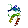
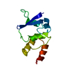
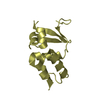
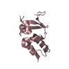
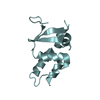
| Title | CRYSTAL STRUCTURE OF THE V135R MUTANT OF A SHAKER T1 DOMAIN | ||||||
 Components Components | POTASSIUM CHANNEL KV1.1 | ||||||
 Keywords Keywords | MEMBRANE PROTEIN / POTASSIUM CHANNELS / APLYSIA KV1.1 / PROTON TRANSPORT | ||||||
| Function / homology |  Function and homology information Function and homology informationdelayed rectifier potassium channel activity / action potential / voltage-gated potassium channel complex / protein homooligomerization Similarity search - Function | ||||||
| Biological species |  | ||||||
| Method |  X-RAY DIFFRACTION / SYNCHROTRON / Resolution: 1.704 Å X-RAY DIFFRACTION / SYNCHROTRON / Resolution: 1.704 Å | ||||||
 Authors Authors | Nanao, M.H. / Cushman, S.J. / Jahng, A.W. / DeRubeis, D. / Choe, S. / Pfaffinger, P.J. | ||||||
 Citation Citation | Journal: Nat.Struct.Biol. / Year: 2000 Title: Voltage dependent activation of potassium channels is coupled to T1 domain structure. Authors: Cushman, S.J. / Nanao, M.H. / Jahng, A.W. / DeRubeis, D. / Choe, S. / Pfaffinger, P.J. | ||||||
| History |
|
- Structure visualization
| Structure viewer | Molecule: MolmilJmol/JSmol |
|---|
- Downloads & links
-Download
| PDBx/mmCIF format | 1eoe.cif.gz | 54.2 KB | Display | PDBx/mmCIF format |
|---|---|---|---|---|
| PDB format | pdb1eoe.ent.gz | 40.4 KB | Display | PDB format |
| PDBx/mmJSON format | 1eoe.json.gz | Tree view | PDBx/mmJSON format | |
| Others |  Other downloads Other downloads |
-Validation report
| Summary document | 1eoe_validation.pdf.gz | 360.9 KB | Display | wwPDB validaton report |
|---|---|---|---|---|
| Full document | 1eoe_full_validation.pdf.gz | 363.8 KB | Display | |
| Data in XML | 1eoe_validation.xml.gz | 3.6 KB | Display | |
| Data in CIF | 1eoe_validation.cif.gz | 5.6 KB | Display | |
| Arichive directory | https://data.pdbj.org/pub/pdb/validation_reports/eo/1eoeftp://data.pdbj.org/pub/pdb/validation_reports/eo/1eoe | HTTPS FTP |
-Related structure data


-Links
 PDBj
PDBj


- Assembly
| Deposited unit | 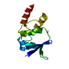
| ||||||||
|---|---|---|---|---|---|---|---|---|---|
| 1 |
| ||||||||
| Unit cell |
|
-Components
| #1: Protein | Mass: 12155.516 Da / Num. of mol.: 1 / Fragment: SHAKER T1 DOMAIN / Mutation: V135R Source method: isolated from a genetically manipulated source Source: (gene. exp.) Plasmid: PET20 / Species (production host): Escherichia coli / Production host:  |
|---|---|
| #2: Water | ChemComp-HOH /  Mass: 18.015 Da / Num. of mol.: 103 / Source method: isolated from a natural source / Formula: H2O Mass: 18.015 Da / Num. of mol.: 103 / Source method: isolated from a natural source / Formula: H2O |
-Experimental details
-Experiment
| Experiment | Method: X-RAY DIFFRACTION / Number of used crystals: 1 |
|---|
- Sample preparation
| Crystal | Density Matthews: 2.73 Å3/Da / Density % sol: 54.92 % | ||||||||||||||||||||||||||||||
|---|---|---|---|---|---|---|---|---|---|---|---|---|---|---|---|---|---|---|---|---|---|---|---|---|---|---|---|---|---|---|---|
| Crystal grow | Temperature: 277 K / Method: vapor diffusion, hanging drop / pH: 7.5 Details: 30% Isopropanol, .1 M Hepes 7.5, .2 M MgCl2, VAPOR DIFFUSION, HANGING DROP, temperature 4K | ||||||||||||||||||||||||||||||
| Crystal grow | *PLUS Method: vapor diffusion / Details: Bixby, K.A., (1999) Nature Struct. Biol., 6, 38. | ||||||||||||||||||||||||||||||
| Components of the solutions | *PLUS
|
-Data collection
| Diffraction | Mean temperature: 100 K |
|---|---|
| Diffraction source | Source: SYNCHROTRON / Site: ALS  / Beamline: 5.0.2 / Wavelength: 1 / Beamline: 5.0.2 / Wavelength: 1 |
| Detector | Type: ADSC QUANTUM 4 / Detector: CCD / Date: Sep 27, 1999 |
| Radiation | Protocol: SINGLE WAVELENGTH / Monochromatic (M) / Laue (L): M / Scattering type: x-ray |
| Radiation wavelength | Wavelength: 1 Å / Relative weight: 1 |
| Reflection | Resolution: 1.7→20 Å / Num. all: 15106 / Num. obs: 15106 / % possible obs: 92.2 % / Observed criterion σ(F): 0 / Observed criterion σ(I): 0 / Redundancy: 8 % / Biso Wilson estimate: 20.316 Å2 / Rmerge(I) obs: 0.078 / Net I/σ(I): 13.5 |
| Reflection shell | Resolution: 1.7→1.73 Å / Redundancy: 2.5 % / Rmerge(I) obs: 0.234 / % possible all: 80.7 |
| Reflection | *PLUS Num. measured all: 130025 |
- Processing
| Software |
| ||||||||||||||||||||
|---|---|---|---|---|---|---|---|---|---|---|---|---|---|---|---|---|---|---|---|---|---|
| Refinement | Resolution: 1.704→19.84 Å / σ(F): 0 / σ(I): 0 / Stereochemistry target values: KONNERT-HENDRICKSON
| ||||||||||||||||||||
| Refinement step | Cycle: LAST / Resolution: 1.704→19.84 Å
| ||||||||||||||||||||
| Software | *PLUS Name: REFMAC / Classification: refinement | ||||||||||||||||||||
| Refinement | *PLUS σ(F): 0 | ||||||||||||||||||||
| Solvent computation | *PLUS | ||||||||||||||||||||
| Displacement parameters | *PLUS Biso mean: 22.5 Å2 | ||||||||||||||||||||
| Refine LS restraints | *PLUS
|
