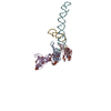
+ Open data
- Basic information
| Entry | Database: PDB / ID: 1emi | ||||||
|---|---|---|---|---|---|---|---|
| Title | STRUCTURE OF 16S RRNA IN THE REGION AROUND RIBOSOMAL PROTEIN S8. | ||||||
 Components Components |
| ||||||
 Keywords Keywords | RIBOSOME / RNA / rRNA / ribosomal protein / 16S rRNA / S8 | ||||||
| Function / homology |  Function and homology information Function and homology informationrRNA binding / structural constituent of ribosome / ribosome / translation / ribonucleoprotein complex / cytoplasm Similarity search - Function | ||||||
| Biological species |   Thermus thermophilus (bacteria) Thermus thermophilus (bacteria) | ||||||
| Method |  X-RAY DIFFRACTION / X-RAY DIFFRACTION /  SYNCHROTRON / Resolution: 7.5 Å SYNCHROTRON / Resolution: 7.5 Å | ||||||
 Authors Authors | Lancaster, L. / Culver, G.M. / Yusupova, G.Z. / Cate, J.H. / Yuspov, M.M. / Noller, H.F. | ||||||
 Citation Citation |  Journal: RNA / Year: 2000 Journal: RNA / Year: 2000Title: The location of protein S8 and surrounding elements of 16S rRNA in the 70S ribosome from combined use of directed hydroxyl radical probing and X-ray crystallography. Authors: Lancaster, L. / Culver, G.M. / Yusupova, G.Z. / Cate, J.H. / Yusupov, M.M. / Noller, H.F. | ||||||
| History |
|
- Structure visualization
| Structure viewer | Molecule:  Molmil Molmil Jmol/JSmol Jmol/JSmol |
|---|
- Downloads & links
- Download
| PDBx/mmCIF format |  1emi.cif.gz 1emi.cif.gz | 22.9 KB | Display |  PDBx/mmCIF format PDBx/mmCIF format |
|---|---|---|---|---|
| PDB format |  pdb1emi.ent.gz pdb1emi.ent.gz | 10.5 KB | Display |  PDB format PDB format |
| PDBx/mmJSON format |  1emi.json.gz 1emi.json.gz | Tree view |  PDBx/mmJSON format PDBx/mmJSON format | |
| Others |  Other downloads Other downloads |
-Validation report
| Arichive directory |  https://data.pdbj.org/pub/pdb/validation_reports/em/1emi https://data.pdbj.org/pub/pdb/validation_reports/em/1emi ftp://data.pdbj.org/pub/pdb/validation_reports/em/1emi ftp://data.pdbj.org/pub/pdb/validation_reports/em/1emi | HTTPS FTP |
|---|
-Related structure data
| Related structure data | |
|---|---|
| Similar structure data |
- Links
Links
- Assembly
| Deposited unit | 
| ||||||||||
|---|---|---|---|---|---|---|---|---|---|---|---|
| 1 |
| ||||||||||
| Unit cell |
|
- Components
| #1: RNA chain | Mass: 51921.684 Da / Num. of mol.: 1 / Source method: isolated from a natural source Details: COORDINATES FOR THERMUS THERMOPHILUS 16S RRNA BUT SEQUENCE AND NUMBERING IS THAT OF E.COLI 16S RRNA Source: (natural)   Thermus thermophilus (bacteria) Thermus thermophilus (bacteria) |
|---|---|
| #2: Protein | Mass: 15624.214 Da / Num. of mol.: 1 / Source method: isolated from a natural source / Source: (natural)   Thermus thermophilus (bacteria) / References: UniProt: Q5SHQ2, UniProt: P0DOY9*PLUS Thermus thermophilus (bacteria) / References: UniProt: Q5SHQ2, UniProt: P0DOY9*PLUS |
-Experimental details
-Experiment
| Experiment | Method:  X-RAY DIFFRACTION / Number of used crystals: 5 X-RAY DIFFRACTION / Number of used crystals: 5 |
|---|
- Sample preparation
| Crystal grow | *PLUS Method: unknown |
|---|
-Data collection
| Diffraction | Mean temperature: 105 K |
|---|---|
| Diffraction source | Source:  SYNCHROTRON / Site: SYNCHROTRON / Site:  ALS ALS  / Type: / Type:  ALS ALS  / Wavelength: 1.1051 / Wavelength: 1.1051 |
| Detector | Type: ADSC QUANTUM 4 / Detector: CCD |
| Radiation | Protocol: SINGLE WAVELENGTH / Monochromatic (M) / Laue (L): M / Scattering type: x-ray |
| Radiation wavelength | Wavelength: 1.1051 Å / Relative weight: 1 |
| Reflection | Highest resolution: 7.5 Å |
- Processing
| Software |
| ||||||||||||
|---|---|---|---|---|---|---|---|---|---|---|---|---|---|
| Refinement | Highest resolution: 7.5 Å Details: All data collection parameters are as reported for 486D. The RNA model for the S8 region of 16S rRNA was built manually by fitting to the 7.8 A X-ray map of the Thermus thermophilus 70S ...Details: All data collection parameters are as reported for 486D. The RNA model for the S8 region of 16S rRNA was built manually by fitting to the 7.8 A X-ray map of the Thermus thermophilus 70S ribosome (Cate et al., Science 285, 2095-2104, 1999; see PDB 486D) using a single P pseudoatom for each nucleotide. Note that the nucleotide numbering corresponds to the nucleotide positions of E.coli 16S rRNA. Protein S8 from Thermus thermophilus (PDB 1AN7) was fitted to the 7.8 A map of the Thermus thermophilus 70S ribosome by splitting the protein into N- and C-terminal domains between residues 80 and 81. The two domains were fit separately, resulting in a small relative movement compared with the original X-ray structure. Only C-alpha positions are given in the PDB file. | ||||||||||||
| Refinement step | Cycle: LAST / Highest resolution: 7.5 Å
|
 Movie
Movie Controller


 PDBj
PDBj


