 Movie
Movie Controller

[English] 日本語
 Yorodumi
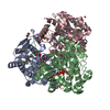
- PDB-1ekk: CRYSTAL STRUCTURE OF HYDROXYETHYLTHIAZOLE KINASE IN THE R3 FORM W... -
+ Open data

- Basic information
| Entry | Database: PDB / ID: 1ekk | ||||||
|---|---|---|---|---|---|---|---|
| Title | CRYSTAL STRUCTURE OF HYDROXYETHYLTHIAZOLE KINASE IN THE R3 FORM WITH HYDROXYETHYLTHIAZOLE | ||||||
 Components Components | HYDROXYETHYLTHIAZOLE KINASE | ||||||
 Keywords Keywords | TRANSFERASE / Alpha-beta | ||||||
| Function / homology |  Function and homology information Function and homology informationhydroxyethylthiazole kinase / hydroxyethylthiazole kinase activity / thiamine biosynthetic process / thiamine diphosphate biosynthetic process / magnesium ion binding / ATP binding Similarity search - Function | ||||||
| Biological species |  | ||||||
| Method |  X-RAY DIFFRACTION / SYNCHROTRON / Resolution: 2 Å X-RAY DIFFRACTION / SYNCHROTRON / Resolution: 2 Å | ||||||
 Authors Authors | Campobasso, N. / Mathews, I.I. / Begley, T.P. / Ealick, S.E. | ||||||
 Citation Citation | Journal: Biochemistry / Year: 2000 Title: Crystal structure of 4-methyl-5-beta-hydroxyethylthiazole kinase from Bacillus subtilis at 1.5 A resolution. Authors: Campobasso, N. / Mathews, I.I. / Begley, T.P. / Ealick, S.E. | ||||||
| History |
|
- Structure visualization
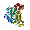
| Structure viewer | Molecule: MolmilJmol/JSmol |
|---|
- Downloads & links
-Download
| PDBx/mmCIF format | 1ekk.cif.gz | 112 KB | Display | PDBx/mmCIF format |
|---|---|---|---|---|
| PDB format | pdb1ekk.ent.gz | 87.1 KB | Display | PDB format |
| PDBx/mmJSON format | 1ekk.json.gz | Tree view | PDBx/mmJSON format | |
| Others |  Other downloads Other downloads |
-Validation report
| Arichive directory | https://data.pdbj.org/pub/pdb/validation_reports/ek/1ekkftp://data.pdbj.org/pub/pdb/validation_reports/ek/1ekk | HTTPS FTP |
|---|
-Related structure data


-Links
 PDBj
PDBj- Assembly
| Deposited unit | 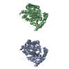
| |||||||||||||||
|---|---|---|---|---|---|---|---|---|---|---|---|---|---|---|---|---|
| 1 | 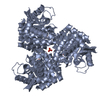
| |||||||||||||||
| 2 | 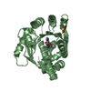
| |||||||||||||||
| 3 | 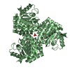
| |||||||||||||||
| Unit cell |
| |||||||||||||||
| Components on special symmetry positions |
| |||||||||||||||
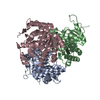
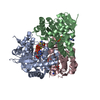
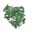
| Details | biological assembly is a trimer. The crystallographic three-fold generates a trimer from chain A and chain B, respectively |
-Components
| #1: Protein | Mass: 28272.166 Da / Num. of mol.: 2 / Mutation: C198(CSD) Source method: isolated from a genetically manipulated source Source: (gene. exp.) #2: Chemical |   Mass: 143.207 Da / Num. of mol.: 2 / Source method: obtained synthetically / Formula: C6H9NOS Mass: 143.207 Da / Num. of mol.: 2 / Source method: obtained synthetically / Formula: C6H9NOS#3: Chemical |   Mass: 64.064 Da / Num. of mol.: 2 / Source method: obtained synthetically / Formula: O2S Mass: 64.064 Da / Num. of mol.: 2 / Source method: obtained synthetically / Formula: O2S#4: Water | ChemComp-HOH / |  Mass: 18.015 Da / Num. of mol.: 182 / Source method: isolated from a natural source / Formula: H2O Mass: 18.015 Da / Num. of mol.: 182 / Source method: isolated from a natural source / Formula: H2OHas protein modification | Y | |
|---|
-Experimental details
-Experiment
| Experiment | Method: X-RAY DIFFRACTION / Number of used crystals: 1 |
|---|
- Sample preparation
| Crystal | Density Matthews: 2.41 Å3/Da / Density % sol: 49.02 % | |||||||||||||||||||||||||
|---|---|---|---|---|---|---|---|---|---|---|---|---|---|---|---|---|---|---|---|---|---|---|---|---|---|---|
| Crystal grow | Temperature: 298 K / Method: vapor diffusion, hanging drop / pH: 7.5 Details: 100mM Tris, 100mM ammonium sulfate, 30% PEG 4000, pH 7.5, VAPOR DIFFUSION, HANGING DROP, temperature 298K | |||||||||||||||||||||||||
| Crystal | *PLUS Density % sol: 49 % | |||||||||||||||||||||||||
| Crystal grow | *PLUS Details: drop consists of equal volume of protein and reservoir solutions | |||||||||||||||||||||||||
| Components of the solutions | *PLUS
|
-Data collection
| Diffraction | Mean temperature: 110 K |
|---|---|
| Diffraction source | Source: SYNCHROTRON / Site: CHESS  / Beamline: F1 / Wavelength: 0.94 / Beamline: F1 / Wavelength: 0.94 |
| Detector | Type: ADSC QUANTUM 4 / Detector: CCD / Date: Nov 25, 1998 |
| Radiation | Protocol: SINGLE WAVELENGTH / Monochromatic (M) / Laue (L): M / Scattering type: x-ray |
| Radiation wavelength | Wavelength: 0.94 Å / Relative weight: 1 |
| Reflection | Resolution: 2→10 Å / Num. all: 35800 / Num. obs: 35426 / % possible obs: 99 % / Observed criterion σ(F): 1 / Observed criterion σ(I): 1 / Redundancy: 5.9 % / Biso Wilson estimate: 20.3 Å2 / Rmerge(I) obs: 0.98 / Net I/σ(I): 17.3 |
| Reflection shell | Resolution: 2→2.1 Å / Redundancy: 3.7 % / Rmerge(I) obs: 0.172 / % possible all: 99 |
| Reflection | *PLUS Num. measured all: 209109 |
| Reflection shell | *PLUS % possible obs: 99 % / Mean I/σ(I) obs: 8.7 |
- Processing
| Software |
| |||||||||||||||||||||||||
|---|---|---|---|---|---|---|---|---|---|---|---|---|---|---|---|---|---|---|---|---|---|---|---|---|---|---|
| Refinement | Resolution: 2→10 Å / σ(F): 0 / σ(I): 0 / Stereochemistry target values: Engh & Huber
| |||||||||||||||||||||||||
| Refinement step | Cycle: LAST / Resolution: 2→10 Å
| |||||||||||||||||||||||||
| Refine LS restraints |
| |||||||||||||||||||||||||
| Software | *PLUS Name: 'CNS' / Classification: refinement | |||||||||||||||||||||||||
| Refine LS restraints | *PLUS
| |||||||||||||||||||||||||
| LS refinement shell | *PLUS Rfactor Rfree: 0.209 / Rfactor Rwork: 0.22 |
