 Movie
Movie Controller

[English] 日本語
 Yorodumi
- PDB-1eh9: CRYSTAL STRUCTURE OF SULFOLOBUS SOLFATARICUS GLYCOSYLTREHALOSE TR... -
+ Open data

- Basic information
| Entry | Database: PDB / ID: 1eh9 | ||||||
|---|---|---|---|---|---|---|---|
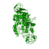
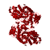
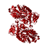
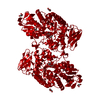
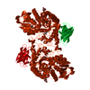
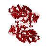
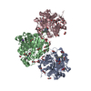
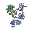
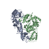
| Title | CRYSTAL STRUCTURE OF SULFOLOBUS SOLFATARICUS GLYCOSYLTREHALOSE TREHALOHYDROLASE | ||||||
 Components Components | GLYCOSYLTREHALOSE TREHALOHYDROLASE | ||||||
 Keywords Keywords | HYDROLASE / trehalose / trehalohydrolase / Sulfolobus solfataricus / alpha/beta barrel / calcium binding / covalent dimer | ||||||
| Function / homology |  Function and homology information Function and homology information4-alpha-D-{(1->4)-alpha-D-glucano}trehalose trehalohydrolase / 4-alpha-D-(1->4)-alpha-D-glucanotrehalose trehalohydrolase activity / trehalose biosynthetic process / cytoplasm Similarity search - Function | ||||||
| Biological species |   Sulfolobus solfataricus (archaea) Sulfolobus solfataricus (archaea) | ||||||
| Method |  X-RAY DIFFRACTION / MIR / Resolution: 3 Å X-RAY DIFFRACTION / MIR / Resolution: 3 Å | ||||||
 Authors Authors | Feese, M.D. / Kato, Y. / Tamada, T. / Kato, M. / Komeda, T. / Kobayashi, K. / Kuroki, R. | ||||||
 Citation Citation | Journal: J.Mol.Biol. / Year: 2000 Title: Crystal structure of glycosyltrehalose trehalohydrolase from the hyperthermophilic archaeum Sulfolobus solfataricus. Authors: Feese, M.D. / Kato, Y. / Tamada, T. / Kato, M. / Komeda, T. / Miura, Y. / Hirose, M. / Hondo, K. / Kobayashi, K. / Kuroki, R. | ||||||
| History |
|
- Structure visualization
| Structure viewer | Molecule: MolmilJmol/JSmol |
|---|
- Downloads & links
-Download
| PDBx/mmCIF format | 1eh9.cif.gz | 125.4 KB | Display | PDBx/mmCIF format |
|---|---|---|---|---|
| PDB format | pdb1eh9.ent.gz | 96.7 KB | Display | PDB format |
| PDBx/mmJSON format | 1eh9.json.gz | Tree view | PDBx/mmJSON format | |
| Others |  Other downloads Other downloads |
-Validation report
| Summary document | 1eh9_validation.pdf.gz | 429.5 KB | Display | wwPDB validaton report |
|---|---|---|---|---|
| Full document | 1eh9_full_validation.pdf.gz | 520.6 KB | Display | |
| Data in XML | 1eh9_validation.xml.gz | 36.2 KB | Display | |
| Data in CIF | 1eh9_validation.cif.gz | 46.9 KB | Display | |
| Arichive directory | https://data.pdbj.org/pub/pdb/validation_reports/eh/1eh9ftp://data.pdbj.org/pub/pdb/validation_reports/eh/1eh9 | HTTPS FTP |
-Related structure data


-Links
 PDBj
PDBj

- Assembly
| Deposited unit | 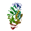
| ||||||||
|---|---|---|---|---|---|---|---|---|---|
| 1 | 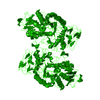
| ||||||||
| Unit cell |
|
-Components
| #1: Protein | Mass: 64741.738 Da / Num. of mol.: 1 Source method: isolated from a genetically manipulated source Source: (gene. exp.) Sulfolobus solfataricus (archaea) / Plasmid: PGUSS2 / Production host:  Pichia jadinii (fungus) / References: UniProt: Q55088, alpha-amylase Pichia jadinii (fungus) / References: UniProt: Q55088, alpha-amylase |
|---|---|
| #2: Water | ChemComp-HOH /  Mass: 18.015 Da / Num. of mol.: 29 / Source method: isolated from a natural source / Formula: H2O Mass: 18.015 Da / Num. of mol.: 29 / Source method: isolated from a natural source / Formula: H2O |
| Has protein modification | Y |
-Experimental details
-Experiment
| Experiment | Method: X-RAY DIFFRACTION / Number of used crystals: 2 |
|---|
- Sample preparation
| Crystal | Density Matthews: 4.1 Å3/Da / Density % sol: 69.7 % | ||||||||||||||||||||
|---|---|---|---|---|---|---|---|---|---|---|---|---|---|---|---|---|---|---|---|---|---|
| Crystal grow | Temperature: 282 K / Method: vapor diffusion, hanging drop / pH: 7.5 Details: 0.6-1.1 M sodium citrate, 0.1 M HEPES, pH 7.5, VAPOR DIFFUSION, HANGING DROP, temperature 282K | ||||||||||||||||||||
| Crystal grow | *PLUS Temperature: 4 ℃ | ||||||||||||||||||||
| Components of the solutions | *PLUS
|
-Data collection
| Diffraction | Mean temperature: 282 K |
|---|---|
| Diffraction source | Source: ROTATING ANODE / Type: RIGAKU RU200 / Wavelength: 1.5418 |
| Detector | Type: RIGAKU RAXIS IIC / Detector: IMAGE PLATE / Date: Jan 1, 1997 / Details: CHARLES SUPPER DOUBLE-MIRROR FOCUSING SYSTEM |
| Radiation | Protocol: SINGLE WAVELENGTH / Monochromatic (M) / Laue (L): M / Scattering type: x-ray |
| Radiation wavelength | Wavelength: 1.5418 Å / Relative weight: 1 |
| Reflection | Resolution: 3→55.9 Å / Num. all: 192375 / Num. obs: 21882 / % possible obs: 96 % / Observed criterion σ(I): -3 / Redundancy: 8.8 % / Biso Wilson estimate: 43.5 Å2 / Rmerge(I) obs: 0.119 / Net I/σ(I): 17.2 |
| Reflection shell | Resolution: 3→3.1 Å / Rmerge(I) obs: 0.873 / Mean I/σ(I) obs: 2.5 / % possible all: 85 |
| Reflection | *PLUS % possible obs: 96 % / Observed criterion σ(F): 2 / Num. measured all: 192375 |
| Reflection shell | *PLUS % possible obs: 85 % |
- Processing
| Software |
| ||||||||||||||||||||||||||||||||||||||||
|---|---|---|---|---|---|---|---|---|---|---|---|---|---|---|---|---|---|---|---|---|---|---|---|---|---|---|---|---|---|---|---|---|---|---|---|---|---|---|---|---|---|
| Refinement | Method to determine structure: MIR / Resolution: 3→20 Å / Stereochemistry target values: Engh & Huber / Details: conjugate direction
| ||||||||||||||||||||||||||||||||||||||||
| Solvent computation | Solvent model: Moews and Kretsinger (1975) J. Mol. Biol., 91: 201-228 Bsol: 686.085 Å2 / ksol: 0.905 e/Å3 | ||||||||||||||||||||||||||||||||||||||||
| Refinement step | Cycle: LAST / Resolution: 3→20 Å
| ||||||||||||||||||||||||||||||||||||||||
| Refine LS restraints |
| ||||||||||||||||||||||||||||||||||||||||
| Software | *PLUS Name: TNT / Version: 5E / Classification: refinement | ||||||||||||||||||||||||||||||||||||||||
| Refinement | *PLUS Highest resolution: 3 Å / Lowest resolution: 20 Å / Num. reflection Rfree: 1116 / % reflection Rfree: 5 % / Rfactor obs: 0.209 / Rfactor Rfree: 0.265 / Rfactor Rwork: 0.203 | ||||||||||||||||||||||||||||||||||||||||
| Solvent computation | *PLUS | ||||||||||||||||||||||||||||||||||||||||
| Displacement parameters | *PLUS | ||||||||||||||||||||||||||||||||||||||||
| Refine LS restraints | *PLUS
| ||||||||||||||||||||||||||||||||||||||||
| LS refinement shell | *PLUS Highest resolution: 3 Å / Lowest resolution: 3.1 Å / Rfactor Rfree: 0.344 / Rfactor obs: 0.264 |
