 Movie
Movie Controller


+ Open data

- Basic information
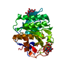
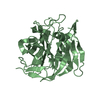
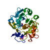
| Entry | Database: PDB / ID: 1egz | ||||||
|---|---|---|---|---|---|---|---|
| Title | CELLULASE CEL5 FROM ERWINIA CHRYSANTHEMI, A FAMILY GH 5-2 ENZYME | ||||||
 Components Components | ENDOGLUCANASE Z | ||||||
 Keywords Keywords | HYDROLASE / GLYCOSYL HYDROLASE / CLAN GH-A / FAMILY 5-2 / CELLULASE | ||||||
| Function / homology |  Function and homology information Function and homology informationcellulase / cellulase activity / cellulose catabolic process / carbohydrate binding / extracellular region Similarity search - Function | ||||||
| Biological species |  Erwinia chrysanthemi (bacteria) Erwinia chrysanthemi (bacteria) | ||||||
| Method |  X-RAY DIFFRACTION / MOLECULAR REPLACEMENT / Resolution: 2.3 Å X-RAY DIFFRACTION / MOLECULAR REPLACEMENT / Resolution: 2.3 Å | ||||||
 Authors Authors | Czjzek, M. / El Hassouni, M. / Py, B. / Barras, F. | ||||||
 Citation Citation | Journal: J.Mol.Biol. / Year: 2001 Title: Type II protein secretion in gram-negative pathogenic bacteria: the study of the structure/secretion relationships of the cellulase Cel5 (formerly EGZ) from Erwinia chrysanthemi Authors: Chapon, M. / Czjzek, M. / El Hassouni, M. / Py, B. / Juy, M. / Barras, F. | ||||||
| History |
|
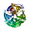
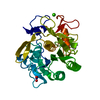
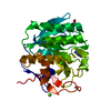
- Structure visualization
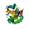
| Structure viewer | Molecule: MolmilJmol/JSmol |
|---|
- Downloads & links
-Download
| PDBx/mmCIF format | 1egz.cif.gz | 185.8 KB | Display | PDBx/mmCIF format |
|---|---|---|---|---|
| PDB format | pdb1egz.ent.gz | 147.2 KB | Display | PDB format |
| PDBx/mmJSON format | 1egz.json.gz | Tree view | PDBx/mmJSON format | |
| Others |  Other downloads Other downloads |
-Validation report
| Summary document | 1egz_validation.pdf.gz | 434.3 KB | Display | wwPDB validaton report |
|---|---|---|---|---|
| Full document | 1egz_full_validation.pdf.gz | 449.3 KB | Display | |
| Data in XML | 1egz_validation.xml.gz | 36.4 KB | Display | |
| Data in CIF | 1egz_validation.cif.gz | 51.3 KB | Display | |
| Arichive directory | https://data.pdbj.org/pub/pdb/validation_reports/eg/1egzftp://data.pdbj.org/pub/pdb/validation_reports/eg/1egz | HTTPS FTP |
-Related structure data
| Related structure data |  1a3hS S: Starting model for refinement |
|---|---|
| Similar structure data |
-Links
 PDBj
PDBj- Assembly
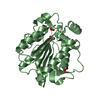
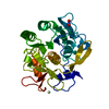
| Deposited unit | 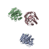
| ||||||||||||
|---|---|---|---|---|---|---|---|---|---|---|---|---|---|
| 1 | 
| ||||||||||||
| 2 | 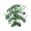
| ||||||||||||
| 3 | 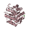
| ||||||||||||
| Unit cell |
| ||||||||||||
| Noncrystallographic symmetry (NCS) | NCS oper:
|
-Components
| #1: Protein | Mass: 32134.514 Da / Num. of mol.: 3 / Fragment: CATALYTIC DOMAIN / Source method: isolated from a natural source / Details: GENE: CELZ / Source: (natural) Erwinia chrysanthemi (bacteria) / Genus: Dickeya / Cellular location: EXTERIOR / Secretion: TYPE II / References: UniProt: P07103, cellulase#2: Chemical |   Mass: 40.078 Da / Num. of mol.: 3 / Source method: obtained synthetically / Formula: Ca Mass: 40.078 Da / Num. of mol.: 3 / Source method: obtained synthetically / Formula: Ca#3: Water | ChemComp-HOH / |  Mass: 18.015 Da / Num. of mol.: 333 / Source method: isolated from a natural source / Formula: H2O Mass: 18.015 Da / Num. of mol.: 333 / Source method: isolated from a natural source / Formula: H2OSequence details | SECRETED (MATURE) PROTEIN, CONSISTING | |
|---|
-Experimental details
-Experiment
| Experiment | Method: X-RAY DIFFRACTION / Number of used crystals: 1 |
|---|
- Sample preparation
| Crystal | Density Matthews: 2.08 Å3/Da / Density % sol: 41 % | ||||||||||||||||||||||||||||||||||||||||||
|---|---|---|---|---|---|---|---|---|---|---|---|---|---|---|---|---|---|---|---|---|---|---|---|---|---|---|---|---|---|---|---|---|---|---|---|---|---|---|---|---|---|---|---|
| Crystal grow | Method: vapor diffusion, hanging drop / pH: 8 Details: VAPOR DIFFUSION HANGING DROP; PEG 4000 28%, TRIS PH 8.0 20 MM, MGSO4 20 MM, VAPOR DIFFUSION, HANGING DROP | ||||||||||||||||||||||||||||||||||||||||||
| Crystal grow | *PLUS Temperature: 4 ℃ / Method: vapor diffusion | ||||||||||||||||||||||||||||||||||||||||||
| Components of the solutions | *PLUS
|
-Data collection
| Diffraction | Mean temperature: 287 K |
|---|---|
| Diffraction source | Source: ROTATING ANODE / Type: RIGAKU RU200 / Wavelength: 1.5418 |
| Detector | Type: MARRESEARCH / Detector: IMAGE PLATE / Date: Dec 15, 1995 |
| Radiation | Monochromator: GRAPHITE SINGLE CRYSTAL / Protocol: SINGLE WAVELENGTH / Monochromatic (M) / Laue (L): M / Scattering type: x-ray |
| Radiation wavelength | Wavelength: 1.5418 Å / Relative weight: 1 |
| Reflection | Resolution: 2.3→29 Å / Num. obs: 38365 / % possible obs: 98 % / Redundancy: 2.8 % / Biso Wilson estimate: 12.3 Å2 / Rsym value: 0.134 / Net I/σ(I): 5 |
| Reflection shell | Resolution: 2.3→2.34 Å / Redundancy: 2.2 % / Mean I/σ(I) obs: 1.5 / Rsym value: 0.45 / % possible all: 97 |
| Reflection | *PLUS Highest resolution: 2.3 Å / % possible obs: 98 % / Rmerge(I) obs: 0.086 |
| Reflection shell | *PLUS Highest resolution: 2.3 Å / % possible obs: 97 % / Num. unique obs: 1749 / Rmerge(I) obs: 0.373 |
- Processing
| Software |
| ||||||||||||||||||||||||||||||||||||||||||||||||||||||||||||
|---|---|---|---|---|---|---|---|---|---|---|---|---|---|---|---|---|---|---|---|---|---|---|---|---|---|---|---|---|---|---|---|---|---|---|---|---|---|---|---|---|---|---|---|---|---|---|---|---|---|---|---|---|---|---|---|---|---|---|---|---|---|
| Refinement | Method to determine structure: MOLECULAR REPLACEMENT Starting model: 1A3H Resolution: 2.3→29 Å / Data cutoff high absF: 100000 / Data cutoff low absF: 0.001 / Cross valid method: THROUGHOUT / σ(F): 0.5
| ||||||||||||||||||||||||||||||||||||||||||||||||||||||||||||
| Displacement parameters | Biso mean: 22.4 Å2 | ||||||||||||||||||||||||||||||||||||||||||||||||||||||||||||
| Refine analyze | Luzzati d res low obs: 29 Å | ||||||||||||||||||||||||||||||||||||||||||||||||||||||||||||
| Refinement step | Cycle: LAST / Resolution: 2.3→29 Å
| ||||||||||||||||||||||||||||||||||||||||||||||||||||||||||||
| Refine LS restraints |
| ||||||||||||||||||||||||||||||||||||||||||||||||||||||||||||
| LS refinement shell | Resolution: 2.3→2.4 Å / Total num. of bins used: 8
| ||||||||||||||||||||||||||||||||||||||||||||||||||||||||||||
| Xplor file | Serial no: 1 / Param file: PARHCSDX.PRO / Topol file: TOPHCSDX.PRO | ||||||||||||||||||||||||||||||||||||||||||||||||||||||||||||
| Refinement | *PLUS Num. reflection obs: 32807 / Rfactor obs: 0.187 / Rfactor Rfree: 0.226 / Rfactor Rwork: 0.187 | ||||||||||||||||||||||||||||||||||||||||||||||||||||||||||||
| Solvent computation | *PLUS | ||||||||||||||||||||||||||||||||||||||||||||||||||||||||||||
| Displacement parameters | *PLUS | ||||||||||||||||||||||||||||||||||||||||||||||||||||||||||||
| Refine LS restraints | *PLUS
| ||||||||||||||||||||||||||||||||||||||||||||||||||||||||||||
| LS refinement shell | *PLUS Rfactor obs: 0.261 |
