[English] 日本語
Yorodumi
- PDB-4gj1: Crystal structure of 1-(5-phosphoribosyl)-5-[(5-phosphoribosylami... -

+
Open data


ID or keywords:

Loading...

-
Basic information

Entry
Database: PDB / ID: 4gj1
TitleCrystal structure of 1-(5-phosphoribosyl)-5-[(5-phosphoribosylamino)methylideneamino] imidazole-4-carboxamide isomerase (hisA).
Components1-(5-phosphoribosyl)-5-[(5-phosphoribosylamino)methylideneamino] imidazole-4-carboxamide isomerase
Keywords
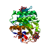
ISOMERASE / HisA / CSGID / NIAID / Structural Genomics / National Institute of Allergy and Infectious Diseases / Center for Structural Genomics of Infectious Diseases / Biosynthesis of secondary metabolites / Histidine metabolism
Function / homology
Function and homology information


1-(5-phosphoribosyl)-5-[(5-phosphoribosylamino)methylideneamino]imidazole-4-carboxamide isomerase / 1-(5-phosphoribosyl)-5-[(5-phosphoribosylamino)methylideneamino]imidazole-4-carboxamide isomerase activity / L-histidine biosynthetic process / cytoplasm
Similarity search - Function
HisA, bacterial-type / HisA/PriA, bacterial-type / Histidine biosynthesis protein / Histidine biosynthesis protein / Ribulose-phosphate binding barrel / Aldolase class I / Aldolase-type TIM barrel / TIM Barrel / Alpha-Beta Barrel / Alpha Beta
Similarity search - Domain/homology
1-(5-phosphoribosyl)-5-[(5-phosphoribosylamino)methylideneamino] imidazole-4-carboxamide isomerase
Similarity search - Component
Biological speciesCampylobacter jejuni subsp. jejuni (Campylobacter)
MethodX-RAY DIFFRACTION / SYNCHROTRON / SAD / Resolution: 2.152 Å
AuthorsNocek, B. / Gu, M. / Kwon, K. / Anderson, W.F. / Joachimiak, A. / CSGID / Center for Structural Genomics of Infectious Diseases (CSGID)
CitationJournal: To be Published
Title: Crystal structure of 1-(5-phosphoribosyl)-5-[(5-phosphoribosylamino)methylideneamino] imidazole-4-carboxamide isomerase (hisA).
Authors: Nocek, B. / Gu, M. / Kwon, K. / Anderson, W.F. / Joachimiak, A.
History
DepositionAug 9, 2012Deposition site: RCSB / Processing site: RCSB
Revision 1.0Aug 22, 2012Provider: repository / Type: Initial release
Revision 1.1Nov 15, 2017Group: Refinement description / Category: software
Revision 1.2Jan 24, 2018Group: Structure summary / Category: audit_author / Item: _audit_author.name

-
Structure visualization

Structure viewerMolecule:
MolmilJmol/JSmol

Downloads & links

-
Assembly

Deposited unit
A: 1-(5-phosphoribosyl)-5-[(5-phosphoribosylamino)methylideneamino] imidazole-4-carboxamide isomerase


Theoretical massNumber of molelcules
Total (without water)26,9091
Polymers26,9091
Non-polymers00
Water77543
1


  • Idetical with deposited unit
  • defined by author&software
TypeNameSymmetry operationNumber
identity operation1_555x,y,z1
MethodPISA
Unit cell
Length a, b, c (Å)35.340, 75.733, 45.923
Angle α, β, γ (deg.)90.00, 105.18, 90.00
Int Tables number4
Space group name H-MP1211

-
Components

#1: Protein 1-(5-phosphoribosyl)-5-[(5-phosphoribosylamino)methylideneamino] imidazole-4-carboxamide isomerase / Phosphoribosylformimino-5-aminoimidazole carboxamide ribotide isomerase


Mass: 26908.908 Da / Num. of mol.: 1
Source method: isolated from a genetically manipulated source
Source: (gene. exp.) Campylobacter jejuni subsp. jejuni (Campylobacter)
Strain: jejuni NCTC 11168 = ATCC 700819 / Gene: hisA, Cj1601 / Plasmid: pMCSG7 / Production host: Escherichia coli (E. coli) / Strain (production host): BL21(DE3)
References: UniProt: Q9PM74, 1-(5-phosphoribosyl)-5-[(5-phosphoribosylamino)methylideneamino]imidazole-4-carboxamide isomerase
#2: Water ChemComp-HOH / water


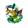
Mass: 18.015 Da / Num. of mol.: 43 / Source method: isolated from a natural source / Formula: H2O

-
Experimental details

-
Experiment

ExperimentMethod: X-RAY DIFFRACTION / Number of used crystals: 1

-
Sample preparation

CrystalDensity Matthews: 2.2 Å3/Da / Density % sol: 44.19 %
Crystal growTemperature: 289 K / Method: vapor diffusion, sitting drop / pH: 7.5
Details: 25% PEG 3350 , 0.2 M lithium Sulfate, pH 7.5, VAPOR DIFFUSION, SITTING DROP, temperature 289K

-
Data collection

DiffractionMean temperature: 100 K
Diffraction sourceSource: SYNCHROTRON / Site: APS / Beamline: 19-ID / Wavelength: 0.9794 Å
DetectorType: ADSC QUANTUM 315r / Detector: CCD / Date: Dec 1, 2011 / Details: mirrors
RadiationMonochromator: double crystal / Protocol: SINGLE WAVELENGTH / Monochromatic (M) / Laue (L): M / Scattering type: x-ray
Radiation wavelengthWavelength: 0.9794 Å / Relative weight: 1
ReflectionResolution: 2.15→35 Å / Num. all: 12600 / Num. obs: 12596 / % possible obs: 99.7 % / Observed criterion σ(F): 2 / Redundancy: 4.1 % / Biso Wilson estimate: 52 Å2 / Rmerge(I) obs: 0.101
Reflection shellResolution: 2.15→2.19 Å / % possible all: 98.6

-
Processing

Software
NameVersionClassification
SBC-Collectdata collection
Auto-Rickshawphasing
PHENIX(phenix.refine: dev_1117)refinement
HKL-3000data reduction
HKL-3000data scaling
RefinementMethod to determine structure: SAD / Resolution: 2.152→34.108 Å / SU ML: 0.29 / σ(F): 2 / σ(I): 0 / Phase error: 25.86 / Stereochemistry target values: ML
RfactorNum. reflection% reflectionSelection details
Rfree0.229 1195 4.93 %RANDOM
Rwork0.1807 ---
all0.19 ---
obs0.183 12576 97.15 %-
Solvent computationShrinkage radii: 0.9 Å / VDW probe radii: 1.11 Å / Solvent model: FLAT BULK SOLVENT MODEL
Refinement stepCycle: LAST / Resolution: 2.152→34.108 Å
ProteinNucleic acidLigandSolventTotal
Num. atoms1714 0 0 43 1757
Refine LS restraints
Refine-IDTypeDev idealNumber
X-RAY DIFFRACTIONf_bond_d0.0041740
X-RAY DIFFRACTIONf_angle_d0.8612344
X-RAY DIFFRACTIONf_dihedral_angle_d13.315640
X-RAY DIFFRACTIONf_chiral_restr0.046288
X-RAY DIFFRACTIONf_plane_restr0.002292
LS refinement shell
Resolution (Å)Rfactor RfreeNum. reflection RfreeRfactor RworkNum. reflection RworkRefine-ID% reflection obs (%)
2.1521-2.23830.33721330.27242196X-RAY DIFFRACTION84
2.2383-2.34010.29321360.24932544X-RAY DIFFRACTION97
2.3401-2.46350.28381220.21832615X-RAY DIFFRACTION99
2.4635-2.61780.2571500.20892596X-RAY DIFFRACTION99
2.6178-2.81980.25941300.20362604X-RAY DIFFRACTION99
2.8198-3.10340.27871080.19852681X-RAY DIFFRACTION100
3.1034-3.55210.28041510.17792612X-RAY DIFFRACTION100
3.5521-4.47360.18261510.15022599X-RAY DIFFRACTION100
4.4736-34.11180.17911140.16212589X-RAY DIFFRACTION98
Refinement TLS params.

Method: refined / Refine-ID: X-RAY DIFFRACTION

IDL112)L122)L132)L222)L232)L332)S11 (Å °)S12 (Å °)S13 (Å °)S21 (Å °)S22 (Å °)S23 (Å °)S31 (Å °)S32 (Å °)S33 (Å °)T112)T122)T132)T222)T232)T332)Origin x (Å)Origin y (Å)Origin z (Å)
17.4198-3.6839-1.42565.45151.47192.62990.2938-0.2030.55280.14260.0651-0.91750.60470.4323-0.36260.3330.101-0.02530.36480.01750.30126.4675-0.79924.5265
29.15141.1341.26519.1478-0.74472.28660.07110.42290.1832-1.4004-0.17041.1147-0.9476-0.8375-0.12350.48950.1431-0.09370.5287-0.03450.4214-6.331311.937322.8748
31.6231.48421.38881.4320.93331.67480.57540.1104-0.8704-0.41540.0592-0.8643-0.23310.4848-0.2790.33310.04020.01110.7109-0.24820.7307-14.41161.98821.1351
46.3617-2.66810.43923.67670.66264.65280.006-0.34911.05090.37870.3593-0.3432-0.1495-0.2692-0.31960.3345-0.0185-0.02740.4042-0.05890.41260.735210.166831.1103
52.7076-1.4015-0.37382.52471.3094.0238-0.0517-0.04330.227-0.08370.2514-0.2583-0.17810.2247-0.19630.26450.0165-0.05140.3970.01670.3464.45126.873622.7775
61.7562-1.57980.40355.14713.66424.45490.59620.84411.0373-0.68080.78640.5481-1.7065-1.6598-0.27910.74710.22990.09490.79780.36890.6375-3.891316.323611.575
76.0356-1.23061.63683.2093-2.33115.35330.4403-0.63240.3854-0.31120.0314-2.3403-1.25090.7372-0.27390.7752-0.09610.16620.3512-0.19160.81438.160216.615722.4937
83.2732-0.4528-1.30254.28620.79523.26250.12590.10230.5455-0.8288-0.1867-0.3741-0.6143-0.5893-0.19670.39410.01660.02040.27550.02340.39455.76138.700812.931
94.533-0.55420.46696.5086-2.27827.03340.2403-0.11081.0709-0.5-0.4257-0.7798-0.65830.34390.03940.5054-0.05380.18550.34220.04270.57799.433911.627711.7937
100.5641-0.3854-0.20010.7303-0.20740.36270.67981.15990.5424-0.52120.43561.2371-0.3961-1.4806-0.01530.4970.0209-0.25550.82990.2190.6425-0.42962.02381.941
117.4484-0.48590.75477.45994.72113.55740.94091.74861.4432-2.0489-0.5415-0.4708-1.2382-0.05880.01590.83880.01390.19790.64150.08830.37229.38645.1914-0.4061
123.79394.2293-3.4047.3294-1.99044.2116-0.546-0.1536-1.4843-0.84720.0106-1.03180.43130.5517-0.01740.4348-0.00720.09460.26030.05070.48539.32630.90869.0289
135.17421.74842.85251.38650.07563.4893-0.01980.3982-0.2132-0.1068-0.36440.3640.8484-0.6047-0.31260.6212-0.2131-0.12140.6091-0.22560.5977-5.3157-12.07027.0363
145.19591.6841-0.23977.4149-0.88344.7948-0.21840.9506-0.7261-1.48620.2154-0.45710.8303-0.7212-0.1380.8323-0.08520.00630.6422-0.06350.34542.6033-9.47231.3484
154.69120.7707-2.9213.5371-0.07277.49680.0047-0.26380.1789-0.44890.2661-0.82660.81690.91060.0390.40610.07340.12450.36860.00560.470411.1183-4.43896.5598
164.09422.50531.18163.48471.34256.9369-1.03550.588-0.971-0.07290.97721.58721.3747-0.41420.05680.7964-0.01020.08740.41360.1130.9333-5.5625-8.309815.0505
176.230.2559-0.39724.5517-0.12144.47790.13230.1022-0.81150.0163-0.29950.01810.9763-0.11340.02070.6631-0.02010.07510.3186-0.08990.45175.7363-14.973213.3003
181.03080.3161-1.18351.05360.08734.5772-0.1217-0.2492-0.18150.61290.16240.22871.1213-0.35030.12290.6027-0.0090.10550.3480.01040.36812.09-8.661523.2508
192.4629-1.1309-0.9966.49973.13544.2661-0.2670.1102-0.31830.8221-0.08270.42340.7439-0.23950.36520.4635-0.00310.00210.39070.00270.32272.3297-6.368325.7881
200.73260.2624-1.00181.31710.83762.6192-0.813-1.0685-0.21061.13440.9676-0.75661.19420.68760.08190.85110.33-0.07750.7192-0.00630.39317.8866-6.08633.2456
Refinement TLS group
IDRefine-IDRefine TLS-IDSelection details
1X-RAY DIFFRACTION1(chain A and resid 2:8)
2X-RAY DIFFRACTION2(chain A and resid 9:16)
3X-RAY DIFFRACTION3(chain A and resid 17:28)
4X-RAY DIFFRACTION4(chain A and resid 29:40)
5X-RAY DIFFRACTION5(chain A and resid 41:56)
6X-RAY DIFFRACTION6(chain A and resid 57:64)
7X-RAY DIFFRACTION7(chain A and resid 65:75)
8X-RAY DIFFRACTION8(chain A and resid 76:88)
9X-RAY DIFFRACTION9(chain A and resid 89:102)
10X-RAY DIFFRACTION10(chain A and resid 103:109)
11X-RAY DIFFRACTION11(chain A and resid 110:121)
12X-RAY DIFFRACTION12(chain A and resid 122:130)
13X-RAY DIFFRACTION13(chain A and resid 131:141)
14X-RAY DIFFRACTION14(chain A and resid 142:160)
15X-RAY DIFFRACTION15(chain A and resid 161:169)
16X-RAY DIFFRACTION16(chain A and resid 170:175)
17X-RAY DIFFRACTION17(chain A and resid 181:197)
18X-RAY DIFFRACTION18(chain A and resid 198:211)
19X-RAY DIFFRACTION19(chain A and resid 212:231)
20X-RAY DIFFRACTION20(chain A and resid 232:243)

+
About Yorodumi

-
News

-
Feb 9, 2022. New format data for meta-information of EMDB entries

New format data for meta-information of EMDB entries

  • Version 3 of the EMDB header file is now the official format.
  • The previous official version 1.9 will be removed from the archive.

Related info.:EMDB header

External links:wwPDB to switch to version 3 of the EMDB data model

-
Aug 12, 2020. Covid-19 info

Covid-19 info

URL: https://pdbjlvh1.pdbj.org/emnavi/covid19.php

New page: Covid-19 featured information page in EM Navigator.

Related info.:Covid-19 info / Mar 5, 2020. Novel coronavirus structure data

+
Mar 5, 2020. Novel coronavirus structure data

Novel coronavirus structure data

Related info.:Yorodumi Speices / Aug 12, 2020. Covid-19 info

External links:COVID-19 featured content - PDBj / Molecule of the Month (242):Coronavirus Proteases

+
Jan 31, 2019. EMDB accession codes are about to change! (news from PDBe EMDB page)

EMDB accession codes are about to change! (news from PDBe EMDB page)

  • The allocation of 4 digits for EMDB accession codes will soon come to an end. Whilst these codes will remain in use, new EMDB accession codes will include an additional digit and will expand incrementally as the available range of codes is exhausted. The current 4-digit format prefixed with “EMD-” (i.e. EMD-XXXX) will advance to a 5-digit format (i.e. EMD-XXXXX), and so on. It is currently estimated that the 4-digit codes will be depleted around Spring 2019, at which point the 5-digit format will come into force.
  • The EM Navigator/Yorodumi systems omit the EMD- prefix.

Related info.:Q: What is EMD? / ID/Accession-code notation in Yorodumi/EM Navigator

External links:EMDB Accession Codes are Changing Soon! / Contact to PDBj

+
Jul 12, 2017. Major update of PDB

Major update of PDB

  • wwPDB released updated PDB data conforming to the new PDBx/mmCIF dictionary.
  • This is a major update changing the version number from 4 to 5, and with Remediation, in which all the entries are updated.
  • In this update, many items about electron microscopy experimental information are reorganized (e.g. em_software).
  • Now, EM Navigator and Yorodumi are based on the updated data.

External links:wwPDB Remediation / Enriched Model Files Conforming to OneDep Data Standards Now Available in the PDB FTP Archive

-
Yorodumi

Thousand views of thousand structures

  • Yorodumi is a browser for structure data from EMDB, PDB, SASBDB, etc.
  • This page is also the successor to EM Navigator detail page, and also detail information page/front-end page for Omokage search.
  • The word "yorodu" (or yorozu) is an old Japanese word meaning "ten thousand". "mi" (miru) is to see.

Related info.:EMDB / PDB / SASBDB / Comparison of 3 databanks / Yorodumi Search / Aug 31, 2016. New EM Navigator & Yorodumi / Yorodumi Papers / Jmol/JSmol / Function and homology information / Changes in new EM Navigator and Yorodumi

Read more