 Movie
Movie Controller


+ Open data

- Basic information
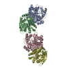
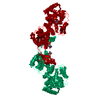
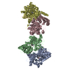
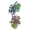
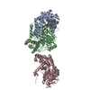
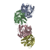
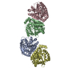
| Entry | Database: PDB / ID: 1eg1 | |||||||||
|---|---|---|---|---|---|---|---|---|---|---|
| Title | ENDOGLUCANASE I FROM TRICHODERMA REESEI | |||||||||
 Components Components | ENDOGLUCANASE I | |||||||||
 Keywords Keywords | CELLULOSE DEGRADATION / ENDOGLUCANASE / MUTATION | |||||||||
| Function / homology |  Function and homology information Function and homology informationcellulose binding / cellulase / cellulase activity / cellulose catabolic process / extracellular region Similarity search - Function | |||||||||
| Biological species |  Hypocrea jecorina (fungus) Hypocrea jecorina (fungus) | |||||||||
| Method |  X-RAY DIFFRACTION / SYNCHROTRON / MOLECULAR REPLACEMENT / Resolution: 3.6 Å X-RAY DIFFRACTION / SYNCHROTRON / MOLECULAR REPLACEMENT / Resolution: 3.6 Å | |||||||||
 Authors Authors | Kleywegt, G.J. / Zou, J.-Y. / Jones, T.A. | |||||||||
 Citation Citation | Journal: J.Mol.Biol. / Year: 1997 Title: The crystal structure of the catalytic core domain of endoglucanase I from Trichoderma reesei at 3.6 A resolution, and a comparison with related enzymes. Authors: Kleywegt, G.J. / Zou, J.Y. / Divne, C. / Davies, G.J. / Sinning, I. / Stahlberg, J. / Reinikainen, T. / Srisodsuk, M. / Teeri, T.T. / Jones, T.A. #1: Journal: J.Mol.Biol. / Year: 1993Title: Crystallization and Preliminary X-Ray Studies on the Core Proteins of Cellobiohydrolase I and Endoglucanase I from Trichoderma Reesei Authors: Divne, C. / Sinning, I. / Stahlberg, J. / Pettersson, G. / Bailey, M. / Siika-Aho, M. / Margolles-Clark, E. / Teeri, T. / Jones, T.A. | |||||||||
| History |
|
- Structure visualization
| Structure viewer | Molecule: MolmilJmol/JSmol |
|---|
- Downloads & links
-Download
| PDBx/mmCIF format | 1eg1.cif.gz | 135.1 KB | Display | PDBx/mmCIF format |
|---|---|---|---|---|
| PDB format | pdb1eg1.ent.gz | 109.1 KB | Display | PDB format |
| PDBx/mmJSON format | 1eg1.json.gz | Tree view | PDBx/mmJSON format | |
| Others |  Other downloads Other downloads |
-Validation report
| Arichive directory | https://data.pdbj.org/pub/pdb/validation_reports/eg/1eg1ftp://data.pdbj.org/pub/pdb/validation_reports/eg/1eg1 | HTTPS FTP |
|---|
-Related structure data
| Similar structure data |
|---|


-Links
 PDBj
PDBj- Assembly
| Deposited unit | 
| ||||||||
|---|---|---|---|---|---|---|---|---|---|
| 1 | 
| ||||||||
| Unit cell |
| ||||||||
| Noncrystallographic symmetry (NCS) | NCS oper: (Code: given Matrix: (-0.999932, -0.01124, 0.003167), Vector: |
-Components
| #1: Protein | Mass: 39169.566 Da / Num. of mol.: 2 / Fragment: CATALYTIC CORE DOMAIN, RESIDUES 1 - 371 / Mutation: DEL(372 - 436) / Source method: isolated from a natural source / Source: (natural) Hypocrea jecorina (fungus) / Plasmid: PEM-F5 / Variant: VTT-D-80133 / Strain: QM9414 / References: UniProt: P07981, cellulase#2: Sugar | ChemComp-NAG /   Type: D-saccharide, beta linking / Mass: 221.208 Da / Num. of mol.: 4 Type: D-saccharide, beta linking / Mass: 221.208 Da / Num. of mol.: 4Source method: isolated from a genetically manipulated source Formula: C8H15NO6 Has protein modification | Y | |
|---|
-Experimental details
-Experiment
| Experiment | Method: X-RAY DIFFRACTION / Number of used crystals: 2 |
|---|
- Sample preparation
| Crystal | Density Matthews: 3.3 Å3/Da / Density % sol: 63 % | |||||||||||||||||||||||||
|---|---|---|---|---|---|---|---|---|---|---|---|---|---|---|---|---|---|---|---|---|---|---|---|---|---|---|
| Crystal grow | pH: 4.5 / Details: DESCRIBED IN REFERENCE 1., pH 4.5 | |||||||||||||||||||||||||
| Crystal grow | *PLUS Method: vapor diffusion, hanging dropDetails: drop contains equal volume of protein and reservoir solutions | |||||||||||||||||||||||||
| Components of the solutions | *PLUS
|
-Data collection
| Diffraction | Mean temperature: 278 K |
|---|---|
| Diffraction source | Source: SYNCHROTRON / Site: EMBL/DESY, HAMBURG  / Beamline: X11 / Beamline: X11 |
| Detector | Type: MARRESEARCH / Detector: IMAGE PLATE / Date: Nov 1, 1992 |
| Radiation | Monochromatic (M) / Laue (L): M / Scattering type: x-ray |
| Radiation wavelength | Relative weight: 1 |
| Reflection | Resolution: 3.6→71.1 Å / Num. obs: 11696 / % possible obs: 92.7 % / Redundancy: 3.9 % / Rmerge(I) obs: 0.117 |
| Reflection shell | Resolution: 3.6→3.69 Å / Redundancy: 4 % / Rmerge(I) obs: 0.404 / % possible all: 93.3 |
| Reflection | *PLUS Num. measured all: 45852 |
| Reflection shell | *PLUS % possible obs: 93.3 % / Num. unique obs: 3353 |
- Processing
| Software |
| ||||||||||||||||||||||||||||||||||||||||||||||||||||||||||||
|---|---|---|---|---|---|---|---|---|---|---|---|---|---|---|---|---|---|---|---|---|---|---|---|---|---|---|---|---|---|---|---|---|---|---|---|---|---|---|---|---|---|---|---|---|---|---|---|---|---|---|---|---|---|---|---|---|---|---|---|---|---|
| Refinement | Method to determine structure: MOLECULAR REPLACEMENT Starting model: HUMICOLA INSOLENS EG1 Resolution: 3.6→8 Å / Cross valid method: THROUGHOUT / σ(F): 0 / Details: GROUPED TEMPERATURE-FACTORS WERE USED.
| ||||||||||||||||||||||||||||||||||||||||||||||||||||||||||||
| Displacement parameters | Biso mean: 35 Å2 | ||||||||||||||||||||||||||||||||||||||||||||||||||||||||||||
| Refinement step | Cycle: LAST / Resolution: 3.6→8 Å
| ||||||||||||||||||||||||||||||||||||||||||||||||||||||||||||
| Refine LS restraints |
| ||||||||||||||||||||||||||||||||||||||||||||||||||||||||||||
| Refine LS restraints NCS | NCS model details: CONSTRAINED | ||||||||||||||||||||||||||||||||||||||||||||||||||||||||||||
| Software | *PLUS Name: X-PLOR / Version: 4 / Classification: refinement | ||||||||||||||||||||||||||||||||||||||||||||||||||||||||||||
| Refinement | *PLUS | ||||||||||||||||||||||||||||||||||||||||||||||||||||||||||||
| Solvent computation | *PLUS | ||||||||||||||||||||||||||||||||||||||||||||||||||||||||||||
| Displacement parameters | *PLUS | ||||||||||||||||||||||||||||||||||||||||||||||||||||||||||||
| Refine LS restraints | *PLUS
|
