+ Open data
- Basic information
| Entry | Database: PDB / ID: 1efc | ||||||
|---|---|---|---|---|---|---|---|
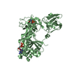
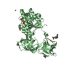
| Title | INTACT ELONGATION FACTOR FROM E.COLI | ||||||
 Components Components | PROTEIN (ELONGATION FACTOR) | ||||||
 Keywords Keywords | RNA BINDING PROTEIN / TRANSPORT AND PROTECTION PROTEIN | ||||||
| Function / homology |  Function and homology information Function and homology informationguanyl-nucleotide exchange factor complex / protein-synthesizing GTPase / guanosine tetraphosphate binding / translational elongation / translation elongation factor activity / response to antibiotic / GTPase activity / GTP binding / magnesium ion binding / RNA binding ...guanyl-nucleotide exchange factor complex / protein-synthesizing GTPase / guanosine tetraphosphate binding / translational elongation / translation elongation factor activity / response to antibiotic / GTPase activity / GTP binding / magnesium ion binding / RNA binding / plasma membrane / cytoplasm Similarity search - Function | ||||||
| Biological species |  | ||||||
| Method |  X-RAY DIFFRACTION / X-RAY DIFFRACTION /  SYNCHROTRON / SYNCHROTRON /  MAD / Resolution: 2.05 Å MAD / Resolution: 2.05 Å | ||||||
 Authors Authors | Song, H. / Parsons, M.R. / Rowsell, S. / Leonard, G. / Phillips, S.E.V. | ||||||
 Citation Citation |  Journal: J.Mol.Biol. / Year: 1999 Journal: J.Mol.Biol. / Year: 1999Title: Crystal structure of intact elongation factor EF-Tu from Escherichia coli in GDP conformation at 2.05 A resolution. Authors: Song, H. / Parsons, M.R. / Rowsell, S. / Leonard, G. / Phillips, S.E. #1:  Journal: Structure / Year: 1996 Journal: Structure / Year: 1996Title: An Alpha to Beta Conformational Switch in EF-TU Authors: Abel, K. / Yoder, M.D. / Hilgenfeld, R. / Jurnak, F. #2:  Journal: Structure / Year: 1996 Journal: Structure / Year: 1996Title: Helix Unwinding in the Effector Region of Elongation Factor EF-TU-Gdp Authors: Polekhina, G. / Thirup, S. / Kjeldgaard, M. / Nissen, P. / Lippmann, C. / Nyborg, J. #3:  Journal: Science / Year: 1995 Journal: Science / Year: 1995Title: Crystal Structure of the Ternary Complex of Phe-tRNA, EF-TU and GTP Analogue Authors: Nissen, P. / Kjeldgaard, M. / Thirup, S. / Polekhina, G. / Reshetnikova, L. / Clark, B.F.C. / Nyborg, J. #4:  Journal: Structure / Year: 1993 Journal: Structure / Year: 1993Title: The Crystal Structure of Elongation Factor EF-TU from T. Aquaticus in the GTP Conformation Authors: Kjeldgaard, M. / Nissen, P. / Thirup, S. / Nyborg, J. #5:  Journal: J.Mol.Biol. / Year: 1992 Journal: J.Mol.Biol. / Year: 1992Title: Refined Structure of Elongation Factor EF-TU from E. Coli Authors: Kjeldgaard, M. / Nyborg, J. | ||||||
| History |
|
- Structure visualization
| Structure viewer | Molecule:  Molmil Molmil Jmol/JSmol Jmol/JSmol |
|---|
- Downloads & links
- Download
| PDBx/mmCIF format |  1efc.cif.gz 1efc.cif.gz | 170.2 KB | Display |  PDBx/mmCIF format PDBx/mmCIF format |
|---|---|---|---|---|
| PDB format |  pdb1efc.ent.gz pdb1efc.ent.gz | 134.6 KB | Display |  PDB format PDB format |
| PDBx/mmJSON format |  1efc.json.gz 1efc.json.gz | Tree view |  PDBx/mmJSON format PDBx/mmJSON format | |
| Others |  Other downloads Other downloads |
-Validation report
| Summary document |  1efc_validation.pdf.gz 1efc_validation.pdf.gz | 510.9 KB | Display |  wwPDB validaton report wwPDB validaton report |
|---|---|---|---|---|
| Full document |  1efc_full_validation.pdf.gz 1efc_full_validation.pdf.gz | 530.5 KB | Display | |
| Data in XML |  1efc_validation.xml.gz 1efc_validation.xml.gz | 18.9 KB | Display | |
| Data in CIF |  1efc_validation.cif.gz 1efc_validation.cif.gz | 30.7 KB | Display | |
| Arichive directory |  https://data.pdbj.org/pub/pdb/validation_reports/ef/1efc https://data.pdbj.org/pub/pdb/validation_reports/ef/1efc ftp://data.pdbj.org/pub/pdb/validation_reports/ef/1efc ftp://data.pdbj.org/pub/pdb/validation_reports/ef/1efc | HTTPS FTP |
-Related structure data
| Similar structure data |
|---|
- Links
Links
- Assembly
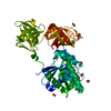
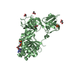
| Deposited unit | 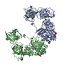
| ||||||||||
|---|---|---|---|---|---|---|---|---|---|---|---|
| 1 | 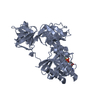
| ||||||||||
| 2 | 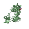
| ||||||||||
| Unit cell |
|
- Components
| #1: Protein | Mass: 43239.297 Da / Num. of mol.: 2 Source method: isolated from a genetically manipulated source Source: (gene. exp.)   #2: Chemical | #3: Chemical | #4: Water | ChemComp-HOH / | Sequence details | EF-TU IS CODED FOR BY TWO DIFFERENT GENES. THE SEQUENCE STRUCTURE ANALYSIS CARRIED OUT ON THIS ...EF-TU IS CODED FOR BY TWO DIFFERENT GENES. THE SEQUENCE STRUCTURE ANALYSIS CARRIED OUT ON THIS MIXTURE SHOWS THAT THE C-TERMINAL RESIDUE OCCURS AS GLY/SER IN THE RATIO OF 3/ 1. THIS RESIDUE IS IDENTIFIED | |
|---|
-Experimental details
-Experiment
| Experiment | Method:  X-RAY DIFFRACTION / Number of used crystals: 4 X-RAY DIFFRACTION / Number of used crystals: 4 |
|---|
- Sample preparation
| Crystal | Density Matthews: 3 Å3/Da / Density % sol: 60 % | ||||||||||||||||||||||||||||||||||||||||||||||||
|---|---|---|---|---|---|---|---|---|---|---|---|---|---|---|---|---|---|---|---|---|---|---|---|---|---|---|---|---|---|---|---|---|---|---|---|---|---|---|---|---|---|---|---|---|---|---|---|---|---|
| Crystal grow | pH: 7.5 / Details: pH 7.5 | ||||||||||||||||||||||||||||||||||||||||||||||||
| Crystal grow | *PLUS pH: 8.5 / Method: vapor diffusion, hanging drop | ||||||||||||||||||||||||||||||||||||||||||||||||
| Components of the solutions | *PLUS
|
-Data collection
| Diffraction | Mean temperature: 100 K | ||||||||||||
|---|---|---|---|---|---|---|---|---|---|---|---|---|---|
| Diffraction source | Source:  SYNCHROTRON / Site: SYNCHROTRON / Site:  ESRF ESRF  / Beamline: BM14 / Wavelength: 0.97899,0.97916,0.91850 / Beamline: BM14 / Wavelength: 0.97899,0.97916,0.91850 | ||||||||||||
| Detector | Type: MARRESEARCH / Detector: IMAGE PLATE / Date: Oct 15, 1996 / Details: MIRRORS | ||||||||||||
| Radiation | Protocol: MAD / Monochromatic (M) / Laue (L): M / Scattering type: x-ray | ||||||||||||
| Radiation wavelength |
| ||||||||||||
| Reflection | Resolution: 2.05→25 Å / Num. obs: 61876 / % possible obs: 90.5 % / Redundancy: 3.5 % / Biso Wilson estimate: 21.8 Å2 / Rmerge(I) obs: 0.082 / Rsym value: 0.082 | ||||||||||||
| Reflection shell | Resolution: 2.05→2.1 Å / Redundancy: 3.5 % / Rmerge(I) obs: 0.274 / % possible all: 91.7 | ||||||||||||
| Reflection | *PLUS Num. measured all: 217915 | ||||||||||||
| Reflection shell | *PLUS % possible obs: 91.7 % |
- Processing
| Software |
| |||||||||||||||||||||||||||||||||||||||||||||||||||||||||||||||
|---|---|---|---|---|---|---|---|---|---|---|---|---|---|---|---|---|---|---|---|---|---|---|---|---|---|---|---|---|---|---|---|---|---|---|---|---|---|---|---|---|---|---|---|---|---|---|---|---|---|---|---|---|---|---|---|---|---|---|---|---|---|---|---|---|
| Refinement | Method to determine structure:  MAD / Resolution: 2.05→10 Å / Cross valid method: THROUGHOUT / ESU R: 0.18 MAD / Resolution: 2.05→10 Å / Cross valid method: THROUGHOUT / ESU R: 0.18
| |||||||||||||||||||||||||||||||||||||||||||||||||||||||||||||||
| Displacement parameters | Biso mean: 24 Å2 | |||||||||||||||||||||||||||||||||||||||||||||||||||||||||||||||
| Refinement step | Cycle: LAST / Resolution: 2.05→10 Å
| |||||||||||||||||||||||||||||||||||||||||||||||||||||||||||||||
| Refine LS restraints |
|
 Movie
Movie Controller





 PDBj
PDBj


