 Movie
Movie Controller

[English] 日本語
 Yorodumi
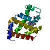
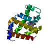
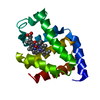
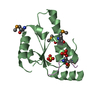
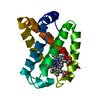
- PDB-1ecd: STRUCTURE OF ERYTHROCRUORIN IN DIFFERENT LIGAND STATES REFINED AT... -
+ Open data

- Basic information
| Entry | Database: PDB / ID: 1ecd | ||||||
|---|---|---|---|---|---|---|---|
| Title | STRUCTURE OF ERYTHROCRUORIN IN DIFFERENT LIGAND STATES REFINED AT 1.4 ANGSTROMS RESOLUTION | ||||||
 Components Components | ERYTHROCRUORIN (AQUO MET) | ||||||
 Keywords Keywords | OXYGEN TRANSPORT | ||||||
| Function / homology |  Function and homology information Function and homology informationhemoglobin complex / oxygen carrier activity / oxygen binding / heme binding / extracellular region / metal ion binding Similarity search - Function | ||||||
| Biological species |  Chironomus thummi thummi (insect) Chironomus thummi thummi (insect) | ||||||
| Method |  X-RAY DIFFRACTION / Resolution: 1.4 Å X-RAY DIFFRACTION / Resolution: 1.4 Å | ||||||
 Authors Authors | Steigemann, W. / Weber, E. | ||||||
 Citation Citation | Journal: J.Mol.Biol. / Year: 1979 Title: Structure of erythrocruorin in different ligand states refined at 1.4 A resolution. Authors: Steigemann, W. / Weber, E. #1: Journal: J.Mol.Biol. / Year: 1978Title: The Structure of Oxy-Erythrocruorin at 1.4 Angstroms Resolution Authors: Weber, E. / Steigemann, W. / Jones, T.A. / Huber, R. #2: Journal: Eur.J.Biochem. / Year: 1971Title: The Atomic Structure of Erythrocruorin in the Light of the Chemical Sequence and its Comparison with Myoglobin Authors: Huber, R. / Epp, O. / Steigemann, W. / Formanek, H. #3: Journal: J.Mol.Biol. / Year: 1970Title: Structures of Deoxy-and Carbomonoxy-Erythrocruorin Authors: Huber, R. / Epp, O. / Formanek, H. | ||||||
| History |
|
- Structure visualization
| Structure viewer | Molecule: MolmilJmol/JSmol |
|---|
- Downloads & links
-Download
| PDBx/mmCIF format | 1ecd.cif.gz | 41.8 KB | Display | PDBx/mmCIF format |
|---|---|---|---|---|
| PDB format | pdb1ecd.ent.gz | 28.3 KB | Display | PDB format |
| PDBx/mmJSON format | 1ecd.json.gz | Tree view | PDBx/mmJSON format | |
| Others |  Other downloads Other downloads |
-Validation report
| Arichive directory | https://data.pdbj.org/pub/pdb/validation_reports/ec/1ecdftp://data.pdbj.org/pub/pdb/validation_reports/ec/1ecd | HTTPS FTP |
|---|
-Related structure data


-Links
 PDBj
PDBj


- Assembly
| Deposited unit | 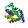
| ||||||||
|---|---|---|---|---|---|---|---|---|---|
| 1 |
| ||||||||
| Unit cell |
| ||||||||
| Atom site foot note | 1: RESIDUE PRO 74 IS A CIS-PROLINE. 2: AN OCCUPANCY OF 0.0 DENOTES ATOMS THAT HAVE NOT BEEN LOCALIZED IN THE ELECTRON DENSITY. 3: THIS HALF OCCUPIED WATER MOLECULE IS THE *SIXTH* LIGAND IN THE REDUCED DEOXY FORM. IT IS LOCATED APPROXIMATELY AT THE SAME POSITION AS THE WATER OF THE AQUO MET FORM 3 ANGSTROMS APART FROM THE IRON. |
-Components
| #1: Protein | Mass: 14800.792 Da / Num. of mol.: 1 Source method: isolated from a genetically manipulated source Source: (gene. exp.) Chironomus thummi thummi (insect) / Species: Chironomus thummi / Strain: thummi / References: UniProt: P02229 |
|---|---|
| #2: Chemical | ChemComp-HEM /   Mass: 616.487 Da / Num. of mol.: 1 / Source method: obtained synthetically / Formula: C34H32FeN4O4 Mass: 616.487 Da / Num. of mol.: 1 / Source method: obtained synthetically / Formula: C34H32FeN4O4 |
| #3: Water | ChemComp-HOH /  Mass: 18.015 Da / Num. of mol.: 95 / Source method: isolated from a natural source / Formula: H2O Mass: 18.015 Da / Num. of mol.: 95 / Source method: isolated from a natural source / Formula: H2O |
-Experimental details
-Experiment
| Experiment | Method: X-RAY DIFFRACTION |
|---|
- Sample preparation
| Crystal | Density Matthews: 2.05 Å3/Da / Density % sol: 39.9 % |
|---|---|
| Crystal grow | *PLUS Method: otherDetails: Braun, V., (1968) Hoppe Seylers Z Physiol Chem., 349, 197. |
-Data collection
| Radiation | Scattering type: x-ray |
|---|---|
| Radiation wavelength | Relative weight: 1 |
| Reflection | *PLUS Highest resolution: 1.45 Å / Num. all: 27170 / Num. obs: 20751 / Rmerge(I) obs: 0.065 |
- Processing
| Refinement | Highest resolution: 1.4 Å | ||||||||||||
|---|---|---|---|---|---|---|---|---|---|---|---|---|---|
| Refinement step | Cycle: LAST / Highest resolution: 1.4 Å
| ||||||||||||
| Refinement | *PLUS Lowest resolution: 7.8 Å / Rfactor obs: 0.183 / Highest resolution: 1.45 Å | ||||||||||||
| Solvent computation | *PLUS | ||||||||||||
| Displacement parameters | *PLUS |
