 Movie
Movie Controller

[English] 日本語
 Yorodumi
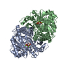
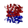
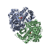
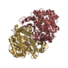
- PDB-1ebh: OCTAHEDRAL COORDINATION AT THE HIGH AFFINITY METAL SITE IN ENOLAS... -
+ Open data

- Basic information
| Entry | Database: PDB / ID: 1ebh | ||||||
|---|---|---|---|---|---|---|---|
| Title | OCTAHEDRAL COORDINATION AT THE HIGH AFFINITY METAL SITE IN ENOLASE; CRYSTALLOGRAPHIC ANALYSIS OF THE MG++-ENZYME FROM YEAST AT 1.9 ANGSTROMS RESOLUTION | ||||||
 Components Components | ENOLASE | ||||||
 Keywords Keywords | CARBON-OXYGEN LYASE | ||||||
| Function / homology |  Function and homology information Function and homology informationGluconeogenesis / regulation of vacuole fusion, non-autophagic / Glycolysis / melatonin binding / phosphopyruvate hydratase / phosphopyruvate hydratase complex / phosphopyruvate hydratase activity / fungal-type vacuole / glycolytic process / magnesium ion binding ...Gluconeogenesis / regulation of vacuole fusion, non-autophagic / Glycolysis / melatonin binding / phosphopyruvate hydratase / phosphopyruvate hydratase complex / phosphopyruvate hydratase activity / fungal-type vacuole / glycolytic process / magnesium ion binding / mitochondrion / plasma membrane / cytoplasm / cytosol Similarity search - Function | ||||||
| Biological species |  | ||||||
| Method |  X-RAY DIFFRACTION / Resolution: 1.9 Å X-RAY DIFFRACTION / Resolution: 1.9 Å | ||||||
 Authors Authors | Wedekind, J.E. / Reed, G.H. / Rayment, I. | ||||||
 Citation Citation | Journal: Biochemistry / Year: 1995 Title: Octahedral coordination at the high-affinity metal site in enolase: crystallographic analysis of the MgII--enzyme complex from yeast at 1.9 A resolution. Authors: Wedekind, J.E. / Reed, G.H. / Rayment, I. #1: Journal: Biochemistry / Year: 1994Title: Chelation of Ser 39 to Mg++ Latches a Gate at the Active Site of Enolase: Structure of the Bis Mg++ Complex of Yeast Enolase and the Intermediate, Phosphonoacetohydroxamate, at 2.1 Angstroms Resolution Authors: Wedekind, J.E. / Reed, G.H. / Rayment, I. | ||||||
| History |
| ||||||
| Remark 700 | SHEET THE SHEETS PRESENTED AS *BAA* AND *BAB* ON SHEET RECORDS BELOW ARE ACTUALLY EIGHT-STRANDED ...SHEET THE SHEETS PRESENTED AS *BAA* AND *BAB* ON SHEET RECORDS BELOW ARE ACTUALLY EIGHT-STRANDED BETA-BARRELS. THESE ARE REPRESENTED BY NINE-STRANDED SHEETS IN WHICH THE FIRST AND LAST STRANDS ARE IDENTICAL. |
- Structure visualization
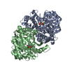
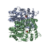
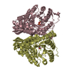
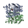
| Structure viewer | Molecule: MolmilJmol/JSmol |
|---|
- Downloads & links
-Download
| PDBx/mmCIF format | 1ebh.cif.gz | 185.7 KB | Display | PDBx/mmCIF format |
|---|---|---|---|---|
| PDB format | pdb1ebh.ent.gz | 146 KB | Display | PDB format |
| PDBx/mmJSON format | 1ebh.json.gz | Tree view | PDBx/mmJSON format | |
| Others |  Other downloads Other downloads |
-Validation report
| Arichive directory | https://data.pdbj.org/pub/pdb/validation_reports/eb/1ebhftp://data.pdbj.org/pub/pdb/validation_reports/eb/1ebh | HTTPS FTP |
|---|
-Related structure data
| Similar structure data |
|---|

-Links
 PDBj
PDBj
- Assembly
| Deposited unit | 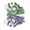
| ||||||||
|---|---|---|---|---|---|---|---|---|---|
| 1 |
| ||||||||
| Unit cell |
| ||||||||
| Atom site foot note | 1: CIS PROLINE - PRO A 143 / 2: CIS PROLINE - PRO B 143 | ||||||||
| Noncrystallographic symmetry (NCS) | NCS oper: (Code: given Matrix: (0.49366, -1.0E-5, -0.86965), Vector: Details | THE TRANSFORMATION PRESENTED ON *MTRIX* RECORDS BELOW WILL YIELD APPROXIMATE COORDINATES FOR CHAIN A WHEN APPLIED TO CHAIN B. THIS TRANSFORMATION IS A TWO-FOLD OPERATION AND TRANSLATION THAT DESCRIBE THE APPROXIMATE NON-CRYSTALLOGRAPHIC DYAD. THE CRYSTALLOGRAPHICALLY INDEPENDENT UNIT IS ONE DIMER OF CHEMICALLY IDENTICAL SUBUNITS. | |
-Components
| #1: Protein | Mass: 46732.797 Da / Num. of mol.: 2 Source method: isolated from a genetically manipulated source Source: (gene. exp.) References: UniProt: P00924, phosphopyruvate hydratase #2: Chemical |   Mass: 35.453 Da / Num. of mol.: 2 / Source method: obtained synthetically / Formula: Cl Mass: 35.453 Da / Num. of mol.: 2 / Source method: obtained synthetically / Formula: Cl#3: Chemical |   Mass: 24.305 Da / Num. of mol.: 2 / Source method: obtained synthetically / Formula: Mg Mass: 24.305 Da / Num. of mol.: 2 / Source method: obtained synthetically / Formula: Mg#4: Water | ChemComp-HOH / |  Mass: 18.015 Da / Num. of mol.: 507 / Source method: isolated from a natural source / Formula: H2O Mass: 18.015 Da / Num. of mol.: 507 / Source method: isolated from a natural source / Formula: H2OCompound details | THIS STRUCTURE COMPLEMENTS THE STUDY OF ENOLASE-BIS(MG)II-PHAH AND SERVES AS A CONTROL EXPERIMENT ...THIS STRUCTURE COMPLEMENT | Nonpolymer details | METALS ARE HEXACOORDINATE WITH OCTAHEDRAL GEOMETRY. A CHLORIDE ION IS BOUND AT THE PHOSPHATE ...METALS ARE HEXACOORDI | |
|---|
-Experimental details
-Experiment
| Experiment | Method: X-RAY DIFFRACTION |
|---|
- Sample preparation
| Crystal | Density Matthews: 2.45 Å3/Da / Density % sol: 49.78 % | ||||||||||||||||||||||||||||||
|---|---|---|---|---|---|---|---|---|---|---|---|---|---|---|---|---|---|---|---|---|---|---|---|---|---|---|---|---|---|---|---|
| Crystal grow | Details: CRYSTALS WERE GROWN FROM POLYETHYLENE GLYCOL, KCL, AT PH 8.1. CRYSTALLIZED IN THE PRESENCE OF 0.5 MILLIMOLAR MG2+ | ||||||||||||||||||||||||||||||
| Crystal grow | *PLUS pH: 8 / Method: batch method | ||||||||||||||||||||||||||||||
| Components of the solutions | *PLUS
|
-Data collection
| Reflection | *PLUS Highest resolution: 1.9 Å / Lowest resolution: 100 Å / Num. obs: 53410 / % possible obs: 75 % / Num. measured all: 77897 / Rmerge(I) obs: 0.041 |
|---|---|
| Reflection shell | *PLUS Highest resolution: 1.9 Å / Lowest resolution: 2.02 Å / Num. unique obs: 6194 / Num. measured obs: 6392 / Rmerge(I) obs: 0.29 |
- Processing
| Software |
| |||||||||||||||||||||||||||||||||||||||||||||||||||||||||||||||
|---|---|---|---|---|---|---|---|---|---|---|---|---|---|---|---|---|---|---|---|---|---|---|---|---|---|---|---|---|---|---|---|---|---|---|---|---|---|---|---|---|---|---|---|---|---|---|---|---|---|---|---|---|---|---|---|---|---|---|---|---|---|---|---|---|
| Refinement | Resolution: 1.9→50 Å / σ(F): 0 Details: OCCUPANCIES FOR RESIDUES EXHIBITING DUAL CONFORMATIONS HAVE TOTAL OCCUPANCIES OF 1.0 WITH THE FOLLOWING EXCEPTION. ALA A 38 AND ALA B 38 WERE MODELED IN THE OPEN CONFORMATION ONL AFTER ATOM ...Details: OCCUPANCIES FOR RESIDUES EXHIBITING DUAL CONFORMATIONS HAVE TOTAL OCCUPANCIES OF 1.0 WITH THE FOLLOWING EXCEPTION. ALA A 38 AND ALA B 38 WERE MODELED IN THE OPEN CONFORMATION ONL AFTER ATOM N ALA 38 IN EACH CHAIN. THE TOTAL OCCUPANCIES FOR ATOMS OF THIS RESIDUE TOTAL 0.7.
| |||||||||||||||||||||||||||||||||||||||||||||||||||||||||||||||
| Refinement step | Cycle: LAST / Resolution: 1.9→50 Å
| |||||||||||||||||||||||||||||||||||||||||||||||||||||||||||||||
| Refine LS restraints |
| |||||||||||||||||||||||||||||||||||||||||||||||||||||||||||||||
| Refinement | *PLUS | |||||||||||||||||||||||||||||||||||||||||||||||||||||||||||||||
| Solvent computation | *PLUS | |||||||||||||||||||||||||||||||||||||||||||||||||||||||||||||||
| Displacement parameters | *PLUS Biso mean: 34 Å2 | |||||||||||||||||||||||||||||||||||||||||||||||||||||||||||||||
| Refine LS restraints | *PLUS
|
