 Movie
Movie Controller

[English] 日本語
 Yorodumi
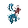
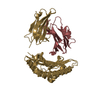
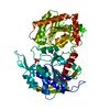
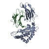
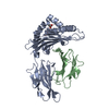
- PDB-1e1n: Structure of adrenodoxin reductase at 2.4 Angstrom in crystal form A' -
+ Open data

- Basic information
| Entry | Database: PDB / ID: 1e1n | ||||||
|---|---|---|---|---|---|---|---|
| Title | Structure of adrenodoxin reductase at 2.4 Angstrom in crystal form A' | ||||||
 Components Components | ADRENODOXIN REDUCTASE | ||||||
 Keywords Keywords | OXIDOREDUCTASE / FLAVOENZYME / ELECTRON TRANSFERASE | ||||||
| Function / homology |  Function and homology information Function and homology informationadrenodoxin-NADP+ reductase / ferredoxin-NADP+ reductase activity / steroid biosynthetic process / ubiquinone biosynthetic process / cholesterol metabolic process / electron transport chain / NADP binding / flavin adenine dinucleotide binding / mitochondrial inner membrane / mitochondrion Similarity search - Function | ||||||
| Biological species |  | ||||||
| Method |  X-RAY DIFFRACTION / MAD / Resolution: 2.4 Å X-RAY DIFFRACTION / MAD / Resolution: 2.4 Å | ||||||
 Authors Authors | Ziegler, G.A. / Schulz, G.E. | ||||||
 Citation Citation | Journal: Biochemistry / Year: 2000 Title: Crystal Structures of Adrenodoxin Reductase in Complex with Nadp+ and Nadph Suggesting a Mechanism for the Electron Transfer of an Enzyme Family Authors: Ziegler, G.A. / Schulz, G.E. #1: Journal: J.Mol.Biol. / Year: 1999Title: The Structure of Adrenodoxin Reductase of Mitochondrial P450 Systems: Electron Transfer for Steroid Biosynthesis Authors: Ziegler, G.A. / Vonrhein, C. / Hanukoglu, I. / Schulz, G.E. #2: Journal: FEBS Lett. / Year: 1999 Title: Chaperone-Assisted Expression of Authentic Bovine Adrenodoxin Reductase in Escherichia Coli Authors: Vonrhein, C. / Schmidt, U. / Ziegler, G.A. / Schweiger, S. / Hanukoglu, I. / Schulz, G.E. | ||||||
| History |
|
- Structure visualization
| Structure viewer | Molecule: MolmilJmol/JSmol |
|---|
- Downloads & links
-Download
| PDBx/mmCIF format | 1e1n.cif.gz | 104.4 KB | Display | PDBx/mmCIF format |
|---|---|---|---|---|
| PDB format | pdb1e1n.ent.gz | 79.2 KB | Display | PDB format |
| PDBx/mmJSON format | 1e1n.json.gz | Tree view | PDBx/mmJSON format | |
| Others |  Other downloads Other downloads |
-Validation report
| Arichive directory | https://data.pdbj.org/pub/pdb/validation_reports/e1/1e1nftp://data.pdbj.org/pub/pdb/validation_reports/e1/1e1n | HTTPS FTP |
|---|
-Related structure data


-Links
 PDBj
PDBj


- Assembly
| Deposited unit | 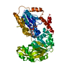
| ||||||||
|---|---|---|---|---|---|---|---|---|---|
| 1 |
| ||||||||
| Unit cell |
|
-Components
| #1: Protein | Mass: 50360.410 Da / Num. of mol.: 1 Source method: isolated from a genetically manipulated source Source: (gene. exp.)  |
|---|---|
| #2: Chemical | ChemComp-FAD /   Mass: 785.550 Da / Num. of mol.: 1 / Source method: obtained synthetically / Formula: C27H33N9O15P2 / Comment: FAD*YM Mass: 785.550 Da / Num. of mol.: 1 / Source method: obtained synthetically / Formula: C27H33N9O15P2 / Comment: FAD*YM |
| #3: Water | ChemComp-HOH /  Mass: 18.015 Da / Num. of mol.: 119 / Source method: isolated from a natural source / Formula: H2O Mass: 18.015 Da / Num. of mol.: 119 / Source method: isolated from a natural source / Formula: H2O |
| Sequence details | FIRST 32 RESIDUES OF SWISS-PROT SEQUENCE REFER TO A MITOCHONDR |
-Experimental details
-Experiment
| Experiment | Method: X-RAY DIFFRACTION / Number of used crystals: 1 |
|---|
- Sample preparation
| Crystal | Density Matthews: 2.82 Å3/Da / Density % sol: 56.42 % | ||||||||||||||||||||||||||||||||||||||||||||||||
|---|---|---|---|---|---|---|---|---|---|---|---|---|---|---|---|---|---|---|---|---|---|---|---|---|---|---|---|---|---|---|---|---|---|---|---|---|---|---|---|---|---|---|---|---|---|---|---|---|---|
| Crystal grow | pH: 6.5 / Details: pH 6.50 | ||||||||||||||||||||||||||||||||||||||||||||||||
| Crystal grow | *PLUS Temperature: 20 ℃ / Method: vapor diffusion, hanging drop | ||||||||||||||||||||||||||||||||||||||||||||||||
| Components of the solutions | *PLUS
|
-Data collection
| Diffraction | Mean temperature: 100 K |
|---|---|
| Diffraction source | Source: ROTATING ANODE / Type: RIGAKU RUH2R / Wavelength: 1.5418 |
| Detector | Type: SIEMENS / Date: Jan 15, 1998 |
| Radiation | Protocol: SINGLE WAVELENGTH / Monochromatic (M) / Laue (L): M / Scattering type: x-ray |
| Radiation wavelength | Wavelength: 1.5418 Å / Relative weight: 1 |
| Reflection | Resolution: 2.35→32.3 Å / Num. obs: 17479 / % possible obs: 74.4 % / Redundancy: 2.4 % / Biso Wilson estimate: 32.8 Å2 / Rmerge(I) obs: 0.057 / Rsym value: 0.048 / Net I/σ(I): 11.4 |
| Reflection shell | Resolution: 2.35→2.48 Å / Redundancy: 1.2 % / Rmerge(I) obs: 0.17 / Mean I/σ(I) obs: 5 / Rsym value: 0.12 / % possible all: 36.8 |
| Reflection shell | *PLUS % possible obs: 36.8 % |
- Processing
| Software | Name: REFMAC / Classification: refinement | |||||||||||||||||||||||||||||||||||||||||||||||||||||||||||||||
|---|---|---|---|---|---|---|---|---|---|---|---|---|---|---|---|---|---|---|---|---|---|---|---|---|---|---|---|---|---|---|---|---|---|---|---|---|---|---|---|---|---|---|---|---|---|---|---|---|---|---|---|---|---|---|---|---|---|---|---|---|---|---|---|---|
| Refinement | Method to determine structure: MAD / Resolution: 2.4→29 Å / σ(F): 0 Details: SMALL DOMAIN MOVEMENT AGAINST OTHER NATIVE STRUCTURES
| |||||||||||||||||||||||||||||||||||||||||||||||||||||||||||||||
| Displacement parameters | Biso mean: 43.1 Å2 | |||||||||||||||||||||||||||||||||||||||||||||||||||||||||||||||
| Refinement step | Cycle: LAST / Resolution: 2.4→29 Å
| |||||||||||||||||||||||||||||||||||||||||||||||||||||||||||||||
| Refine LS restraints |
|
