 Movie
Movie Controller

[English] 日本語
 Yorodumi
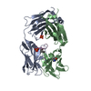
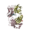
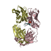
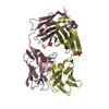
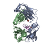
- PDB-1dvr: STRUCTURE OF A MUTANT ADENYLATE KINASE LIGATED WITH AN ATP-ANALOG... -
+ Open data

- Basic information
| Entry | Database: PDB / ID: 1dvr | ||||||
|---|---|---|---|---|---|---|---|
| Title | STRUCTURE OF A MUTANT ADENYLATE KINASE LIGATED WITH AN ATP-ANALOGUE SHOWING DOMAIN CLOSURE OVER ATP | ||||||
 Components Components | ADENYLATE KINASE | ||||||
 Keywords Keywords | TRANSFERASE (PHOSPHOTRANSFERASE) / NUCLEOSIDE MONOPHOSPHATE KINASE / MYOKINASE | ||||||
| Function / homology |  Function and homology information Function and homology informationpre-replicative complex assembly / Interconversion of nucleotide di- and triphosphates / AMP metabolic process / ADP biosynthetic process / adenylate kinase / AMP kinase activity / nucleotide metabolic process / AMP binding / DNA replication origin binding / DNA replication initiation ...pre-replicative complex assembly / Interconversion of nucleotide di- and triphosphates / AMP metabolic process / ADP biosynthetic process / adenylate kinase / AMP kinase activity / nucleotide metabolic process / AMP binding / DNA replication origin binding / DNA replication initiation / ATP metabolic process / mitochondrial intermembrane space / mitochondrion / ATP binding / cytosol / cytoplasm Similarity search - Function | ||||||
| Biological species |  | ||||||
| Method |  X-RAY DIFFRACTION / Resolution: 2.36 Å X-RAY DIFFRACTION / Resolution: 2.36 Å | ||||||
 Authors Authors | Schlauderer, G.J. / Schulz, G.E. | ||||||
 Citation Citation | Journal: J.Mol.Biol. / Year: 1996 Title: Structure of a mutant adenylate kinase ligated with an ATP-analogue showing domain closure over ATP. Authors: Schlauderer, G.J. / Proba, K. / Schulz, G.E. #1: Journal: Eur.J.Biochem. / Year: 1995Title: Stability, Activity and Structure of Adenylate Kinase Mutants Authors: Spuergin, P. / Abele, U. / Schulz, G.E. #2: Journal: Protein Sci. / Year: 1995Title: High-Resolution Structures of Adenylate Kinase from Yeast Ligated with Inhibitor Ap5A, Showing the Pathway of Phosphoryl Transfer Authors: Abele, U. / Schulz, G.E. #3: Journal: Structure / Year: 1995Title: Movie of the Structural Changes During a Catalytic Cycle of Nucleoside Monophosphate Kinases Authors: Vonrhein, C. / Schlauderer, G.J. / Schulz, G.E. #4: Journal: Nucleic Acids Res. / Year: 1987Title: The C-DNA Sequence Encoding Cytosolic Adenylate Kinase from Baker'S Yeast (Saccharomyces Cerevisiae) Authors: Proba, K. / Tomasselli, A.G. / Nielsen, P. / Schulz, G.E. #5: Journal: J.Mol.Biol. / Year: 1987Title: Structure of the Complex of Yeast Adenylate Kinase with the Inhibitor Ap5A at 2.6 Angstroms Resolution Authors: Egner, U. / Tomasselli, A.G. / Schulz, G.E. #6: Journal: Eur.J.Biochem. / Year: 1986Title: The Complete Amino Acid Sequence of Adenylate Kinase from Baker'S Yeast Authors: Tomasselli, A.G. / Mast, E. / Janes, W. / Schiltz, E. | ||||||
| History |
| ||||||
| Remark 700 | SHEET THE HELIX AND SHEET ASSIGNMENTS BELOW ARE AUTOMATIC WITH PROGRAM DSSP. |
- Structure visualization
| Structure viewer | Molecule: MolmilJmol/JSmol |
|---|
- Downloads & links
-Download
| PDBx/mmCIF format | 1dvr.cif.gz | 100.3 KB | Display | PDBx/mmCIF format |
|---|---|---|---|---|
| PDB format | pdb1dvr.ent.gz | 77.6 KB | Display | PDB format |
| PDBx/mmJSON format | 1dvr.json.gz | Tree view | PDBx/mmJSON format | |
| Others |  Other downloads Other downloads |
-Validation report
| Arichive directory | https://data.pdbj.org/pub/pdb/validation_reports/dv/1dvrftp://data.pdbj.org/pub/pdb/validation_reports/dv/1dvr | HTTPS FTP |
|---|
-Related structure data
| Similar structure data |
|---|


-Links
 PDBj
PDBj- Assembly
| Deposited unit | 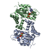
| ||||||||
|---|---|---|---|---|---|---|---|---|---|
| 1 |
| ||||||||
| Unit cell |
| ||||||||
| Atom site foot note | 1: CIS PROLINE - PRO A 92 / 2: CIS PROLINE - PRO B 92 | ||||||||
| Noncrystallographic symmetry (NCS) | NCS oper: (Code: given Matrix: (-0.489945, -0.809503, -0.323511), Vector: |
-Components
| #1: Protein | Mass: 24008.525 Da / Num. of mol.: 2 / Mutation: D89V, R165I Source method: isolated from a genetically manipulated source Source: (gene. exp.) Cellular location: CYTOSOL / Plasmid: PUAKYI / Production host:  #2: Chemical | 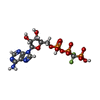  Mass: 541.189 Da / Num. of mol.: 2 / Source method: obtained synthetically / Formula: C11H16F2N5O12P3 Mass: 541.189 Da / Num. of mol.: 2 / Source method: obtained synthetically / Formula: C11H16F2N5O12P3#3: Water | ChemComp-HOH / |  Mass: 18.015 Da / Num. of mol.: 161 / Source method: isolated from a natural source / Formula: H2O Mass: 18.015 Da / Num. of mol.: 161 / Source method: isolated from a natural source / Formula: H2O |
|---|
-Experimental details
-Experiment
| Experiment | Method: X-RAY DIFFRACTION |
|---|
- Sample preparation
| Crystal | Density Matthews: 2.17 Å3/Da / Density % sol: 43.29 % | ||||||||||||||||||||||||||||||||||||||||||||||||||||||
|---|---|---|---|---|---|---|---|---|---|---|---|---|---|---|---|---|---|---|---|---|---|---|---|---|---|---|---|---|---|---|---|---|---|---|---|---|---|---|---|---|---|---|---|---|---|---|---|---|---|---|---|---|---|---|---|
| Crystal | *PLUS Density % sol: 43 % | ||||||||||||||||||||||||||||||||||||||||||||||||||||||
| Crystal grow | *PLUS pH: 6.3 / Method: vapor diffusion, hanging drop | ||||||||||||||||||||||||||||||||||||||||||||||||||||||
| Components of the solutions | *PLUS
|
-Data collection
| Diffraction source | Source: ROTATING ANODE / Type: RIGAKU RU200 / Wavelength: 1.5418 |
|---|---|
| Detector | Type: SIEMENS / Detector: AREA DETECTOR / Date: 1991 |
| Radiation | Monochromatic (M) / Laue (L): M / Scattering type: x-ray |
| Radiation wavelength | Wavelength: 1.5418 Å / Relative weight: 1 |
| Reflection | Resolution: 2.36→10 Å / Num. obs: 15793 / % possible obs: 90 % / Observed criterion σ(I): 0 / Redundancy: 4.1 % / Rmerge(I) obs: 0.058 |
| Reflection | *PLUS Rmerge(I) obs: 0.058 |
- Processing
| Software |
| ||||||||||||||||||||||||||||||||||||||||||||||||||||||||||||||||||||||||||||||||
|---|---|---|---|---|---|---|---|---|---|---|---|---|---|---|---|---|---|---|---|---|---|---|---|---|---|---|---|---|---|---|---|---|---|---|---|---|---|---|---|---|---|---|---|---|---|---|---|---|---|---|---|---|---|---|---|---|---|---|---|---|---|---|---|---|---|---|---|---|---|---|---|---|---|---|---|---|---|---|---|---|---|
| Refinement | Resolution: 2.36→10 Å / σ(F): 0 Details: RESIDUES SER 166 - ASN 169 IN CHAIN HAVE WEAK DENSITY.
| ||||||||||||||||||||||||||||||||||||||||||||||||||||||||||||||||||||||||||||||||
| Displacement parameters | Biso mean: 29 Å2 | ||||||||||||||||||||||||||||||||||||||||||||||||||||||||||||||||||||||||||||||||
| Refine analyze | Luzzati coordinate error obs: 0.28 Å | ||||||||||||||||||||||||||||||||||||||||||||||||||||||||||||||||||||||||||||||||
| Refinement step | Cycle: LAST / Resolution: 2.36→10 Å
| ||||||||||||||||||||||||||||||||||||||||||||||||||||||||||||||||||||||||||||||||
| Refine LS restraints |
| ||||||||||||||||||||||||||||||||||||||||||||||||||||||||||||||||||||||||||||||||
| Software | *PLUS Name: X-PLOR / Classification: refinement | ||||||||||||||||||||||||||||||||||||||||||||||||||||||||||||||||||||||||||||||||
| Refinement | *PLUS | ||||||||||||||||||||||||||||||||||||||||||||||||||||||||||||||||||||||||||||||||
| Solvent computation | *PLUS | ||||||||||||||||||||||||||||||||||||||||||||||||||||||||||||||||||||||||||||||||
| Displacement parameters | *PLUS |
