 Movie
Movie Controller

[English] 日本語
 Yorodumi
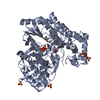
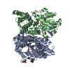
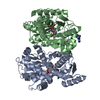
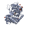
- PDB-1dmu: Crystal structure of the restriction endonuclease BglI (e.c.3.1.2... -
+ Open data

- Basic information
| Entry | Database: PDB / ID: 1dmu | ||||||
|---|---|---|---|---|---|---|---|
| Title | Crystal structure of the restriction endonuclease BglI (e.c.3.1.21.4) bound to its dna recognition sequence | ||||||
 Components Components |
| ||||||
 Keywords Keywords | HYDROLASE/DNA / PROTEIN-DNA COMPLEX / ACTIVE SITE CALCIUM IONS / ALPHA/BETA STRUCTURE / A:A MISMATCH / HYDROLASE-DNA COMPLEX | ||||||
| Function / homology |  Function and homology information Function and homology informationtype II site-specific deoxyribonuclease / type II site-specific deoxyribonuclease activity / DNA restriction-modification system / metal ion binding Similarity search - Function | ||||||
| Biological species |  | ||||||
| Method |  X-RAY DIFFRACTION / SYNCHROTRON / Resolution: 2.2 Å X-RAY DIFFRACTION / SYNCHROTRON / Resolution: 2.2 Å | ||||||
 Authors Authors | Newman, M. / Lunnen, K. / Wilson, G. / Greci, J. / Schildkraut, I. / Phillips, S.E.V. | ||||||
 Citation Citation | Journal: EMBO J. / Year: 1998 Title: Crystal structure of restriction endonuclease BglI bound to its interrupted DNA recognition sequence. Authors: Newman, M. / Lunnen, K. / Wilson, G. / Greci, J. / Schildkraut, I. / Phillips, S.E. #1: Journal: Patent / Year: 1994Title: Method for producing the BglI restriction endonuclease and methylase Authors: Lunnen, K.D. / Wilson, G.G. | ||||||
| History |
|
- Structure visualization
| Structure viewer | Molecule: MolmilJmol/JSmol |
|---|
- Downloads & links
-Download
| PDBx/mmCIF format | 1dmu.cif.gz | 91.9 KB | Display | PDBx/mmCIF format |
|---|---|---|---|---|
| PDB format | pdb1dmu.ent.gz | 65.4 KB | Display | PDB format |
| PDBx/mmJSON format | 1dmu.json.gz | Tree view | PDBx/mmJSON format | |
| Others |  Other downloads Other downloads |
-Validation report
| Arichive directory | https://data.pdbj.org/pub/pdb/validation_reports/dm/1dmuftp://data.pdbj.org/pub/pdb/validation_reports/dm/1dmu | HTTPS FTP |
|---|
-Related structure data
| Similar structure data |
|---|
-Links
 PDBj
PDBj


- Assembly
| Deposited unit | 
| ||||||||||
|---|---|---|---|---|---|---|---|---|---|---|---|
| 1 | 
| ||||||||||
| Unit cell |
|
-Components
| #1: DNA chain | Mass: 5211.398 Da / Num. of mol.: 1 / Source method: obtained synthetically / Details: CONTAINS DNA RECOGNITION SEQUENCE OF BGLI | ||||||
|---|---|---|---|---|---|---|---|
| #2: Protein | Mass: 34050.793 Da / Num. of mol.: 1 Source method: isolated from a genetically manipulated source Source: (gene. exp.) References: UniProt: O68557, type II site-specific deoxyribonuclease | ||||||
| #3: Chemical | ChemComp-CA /   Mass: 40.078 Da / Num. of mol.: 4 / Source method: obtained synthetically / Formula: Ca Mass: 40.078 Da / Num. of mol.: 4 / Source method: obtained synthetically / Formula: Ca#4: Chemical | ChemComp-BME / |   Mass: 78.133 Da / Num. of mol.: 1 / Source method: obtained synthetically / Formula: C2H6OS Mass: 78.133 Da / Num. of mol.: 1 / Source method: obtained synthetically / Formula: C2H6OS#5: Water | ChemComp-HOH / |  Mass: 18.015 Da / Num. of mol.: 255 / Source method: isolated from a natural source / Formula: H2O Mass: 18.015 Da / Num. of mol.: 255 / Source method: isolated from a natural source / Formula: H2OHas protein modification | N | |
-Experimental details
-Experiment
| Experiment | Method: X-RAY DIFFRACTION / Number of used crystals: 1 |
|---|
- Sample preparation
| Crystal | Density Matthews: 2.37 Å3/Da / Density % sol: 48.16 % | ||||||||||||||||||||||||||||||||||||||||||||||||
|---|---|---|---|---|---|---|---|---|---|---|---|---|---|---|---|---|---|---|---|---|---|---|---|---|---|---|---|---|---|---|---|---|---|---|---|---|---|---|---|---|---|---|---|---|---|---|---|---|---|
| Crystal grow | Temperature: 293 K / Method: vapor diffusion, hanging drop / pH: 8.5 Details: 7-12% PEG 4000, 75-150MM LI2SO4, 100MM TRIS-HCL, 1:2 PROTEIN:DNA MOLAR RATIO, pH 8.5, VAPOR DIFFUSION, HANGING DROP, temperature 293K | ||||||||||||||||||||||||||||||||||||||||||||||||
| Components of the solutions |
| ||||||||||||||||||||||||||||||||||||||||||||||||
| Crystal grow | *PLUS pH: 7.5 / Method: vapor diffusion | ||||||||||||||||||||||||||||||||||||||||||||||||
| Components of the solutions | *PLUS
|
-Data collection
| Diffraction | Mean temperature: 100 K |
|---|---|
| Diffraction source | Source: SYNCHROTRON / Site: ESRF  / Beamline: BM14 / Wavelength: 0.919 / Beamline: BM14 / Wavelength: 0.919 |
| Detector | Type: CUSTOM-MADE / Detector: CCD / Date: Jun 11, 1997 |
| Radiation | Protocol: SINGLE WAVELENGTH / Monochromatic (M) / Laue (L): M / Scattering type: x-ray |
| Radiation wavelength | Wavelength: 0.919 Å / Relative weight: 1 |
| Reflection | Resolution: 2.2→50 Å / Num. all: 17679 / Num. obs: 17679 / % possible obs: 90.8 % / Observed criterion σ(F): 0 / Observed criterion σ(I): 0 / Redundancy: 3.3 % / Biso Wilson estimate: 24.3 Å2 / Rmerge(I) obs: 0.055 / Net I/σ(I): 23.3 |
| Reflection shell | Resolution: 2.2→2.25 Å / Redundancy: 0.8 % / Rmerge(I) obs: 0.078 / % possible all: 57.1 |
| Reflection | *PLUS |
| Reflection shell | *PLUS % possible obs: 57.1 % |
- Processing
| Software |
| ||||||||||||||||||||||||||||||||||||||||||||||||||||||||||||
|---|---|---|---|---|---|---|---|---|---|---|---|---|---|---|---|---|---|---|---|---|---|---|---|---|---|---|---|---|---|---|---|---|---|---|---|---|---|---|---|---|---|---|---|---|---|---|---|---|---|---|---|---|---|---|---|---|---|---|---|---|---|
| Refinement | Resolution: 2.2→10 Å / Cross valid method: THROUGHOUT / σ(F): 2 Stereochemistry target values: ENGH & HUBER FOR PROTEIN AND PARKINSON ET AL. FOR DNA Details: TO IMPOSE DOUBLE STRANDED BASE-PAIRING RESTRAINTS ON DNA, TWO OLIGONUCLEOTIDE STRANDS WERE INCLUDED IN THE ASYMMETRIC UNIT (RELATED BY CRYSTALLOGRAPHIC TWO- FOLD). THESE WERE GIVEN 0.5 ...Details: TO IMPOSE DOUBLE STRANDED BASE-PAIRING RESTRAINTS ON DNA, TWO OLIGONUCLEOTIDE STRANDS WERE INCLUDED IN THE ASYMMETRIC UNIT (RELATED BY CRYSTALLOGRAPHIC TWO- FOLD). THESE WERE GIVEN 0.5 OCCUPANCY AND WERE REFINED WITH FORCE CONSTANTS FOR VAN DER WAALS AND ELECTROSTATIC INTERACTIONS BETWEEN SYMMETRY RELATED MOLECULES SET TO ZERO. B-DNA SUGAR PUCKER RESTRAINTS WERE IMPOSED ONLY AT THE BEGINNING OF REFINEMENT. THE FOLLOWING SIDE-CHAINS CANNOT BE POSITIONED IN THE ELECTRON DENSITY MAP: GLU7, GLN15, GLU47, GLU102, GLU172, ARG186, THR224, LYS225 AND LYS259
| ||||||||||||||||||||||||||||||||||||||||||||||||||||||||||||
| Refinement step | Cycle: LAST / Resolution: 2.2→10 Å
| ||||||||||||||||||||||||||||||||||||||||||||||||||||||||||||
| Refine LS restraints |
|
