 Movie
Movie Controller


+ Open data

- Basic information
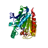
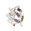
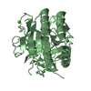
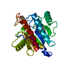
| Entry | Database: PDB / ID: 1din | ||||||
|---|---|---|---|---|---|---|---|
| Title | DIENELACTONE HYDROLASE AT 2.8 ANGSTROMS | ||||||
 Components Components | DIENELACTONE HYDROLASE | ||||||
 Keywords Keywords | HYDROLYTIC ENZYME / DIENELACTONE HYDROLASE / AROMATIC HYDROCARBON CATABOLISM / SERINE ESTERASE / CARBOXYMETHYLENEBUTENOLIDASE | ||||||
| Function / homology |  Function and homology information Function and homology information | ||||||
| Biological species |  Pseudomonas knackmussii (bacteria) Pseudomonas knackmussii (bacteria) | ||||||
| Method |  X-RAY DIFFRACTION / Resolution: 1.8 Å X-RAY DIFFRACTION / Resolution: 1.8 Å | ||||||
 Authors Authors | Ollis, D.L. / Pathak, D. | ||||||
 Citation Citation | Journal: J.Mol.Biol. / Year: 1990 Title: Refined structure of dienelactone hydrolase at 1.8 A. Authors: Pathak, D. / Ollis, D. #1: Journal: J.Mol.Biol. / Year: 1988Title: X-Ray Crystallographic Structure of Dienelactone Hydrolase at 2.8 A Authors: Pathak, D. / Ngai, K.L. / Ollis, D. #2: Journal: J.Biol.Chem. / Year: 1985Title: Crystallization and Preliminary X-Ray Crystallographic Data of Dienelactone Hydrolase from Pseudomonas Sp. B13 Authors: Ollis, D.L. / Ngai, K.L. | ||||||
| History |
|
- Structure visualization
| Structure viewer | Molecule: MolmilJmol/JSmol |
|---|
- Downloads & links
-Download
| PDBx/mmCIF format | 1din.cif.gz | 64.6 KB | Display | PDBx/mmCIF format |
|---|---|---|---|---|
| PDB format | pdb1din.ent.gz | 46.7 KB | Display | PDB format |
| PDBx/mmJSON format | 1din.json.gz | Tree view | PDBx/mmJSON format | |
| Others |  Other downloads Other downloads |
-Validation report
| Arichive directory | https://data.pdbj.org/pub/pdb/validation_reports/di/1dinftp://data.pdbj.org/pub/pdb/validation_reports/di/1din | HTTPS FTP |
|---|
-Related structure data
| Similar structure data |
|---|


-Links
 PDBj
PDBj- Assembly
| Deposited unit | 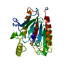
| ||||||||
|---|---|---|---|---|---|---|---|---|---|
| 1 |
| ||||||||
| Unit cell |
|
-Components
| #1: Protein | Mass: 25543.811 Da / Num. of mol.: 1 Source method: isolated from a genetically manipulated source Source: (gene. exp.) Pseudomonas knackmussii (bacteria) / Strain: B13 / Gene: CLC D / Plasmid: PDC100 / Gene (production host): CLC D / Production host: Pseudomonas sp. (bacteria)References: PIR: S02022, UniProt: P0A115*PLUS, carboxymethylenebutenolidase |
|---|---|
| #2: Water | ChemComp-HOH /  Mass: 18.015 Da / Num. of mol.: 279 / Source method: isolated from a natural source / Formula: H2O Mass: 18.015 Da / Num. of mol.: 279 / Source method: isolated from a natural source / Formula: H2O |
| Has protein modification | Y |
| Sequence details | THERE IS MICROHETEROGENEITY AT RESIDUE 123. ONE OF THE RESIDUES IS CYSTEINE AND THE OTHER IS ...THERE IS MICROHETER |
-Experimental details
-Experiment
| Experiment | Method: X-RAY DIFFRACTION |
|---|
- Sample preparation
| Crystal | Density Matthews: 2.67 Å3/Da / Density % sol: 54 % | ||||||||||||||||||||||||
|---|---|---|---|---|---|---|---|---|---|---|---|---|---|---|---|---|---|---|---|---|---|---|---|---|---|
| Crystal grow | *PLUS Temperature: 18 ℃ / pH: 6.3 / Method: vapor diffusion, hanging drop / Details: Pathak, D., (1988) J.Mol.Biol., 204, 435. | ||||||||||||||||||||||||
| Components of the solutions | *PLUS
|
-Data collection
| Diffraction source | Wavelength: 1.5418 |
|---|---|
| Detector | Detector: AREA DETECTOR / Date: 1987 |
| Radiation | Monochromatic (M) / Laue (L): M / Scattering type: x-ray |
| Radiation wavelength | Wavelength: 1.5418 Å / Relative weight: 1 |
| Reflection | Num. obs: 29488 / % possible obs: 95 % / Redundancy: 6 % / Rmerge(I) obs: 0.057 |
- Processing
| Software |
| ||||||||||||||||||||||||||||||||||||||||||||||||||||||||||||||||||||||||||||||||||||
|---|---|---|---|---|---|---|---|---|---|---|---|---|---|---|---|---|---|---|---|---|---|---|---|---|---|---|---|---|---|---|---|---|---|---|---|---|---|---|---|---|---|---|---|---|---|---|---|---|---|---|---|---|---|---|---|---|---|---|---|---|---|---|---|---|---|---|---|---|---|---|---|---|---|---|---|---|---|---|---|---|---|---|---|---|---|
| Refinement | Resolution: 1.8→10 Å / σ(F): 0
| ||||||||||||||||||||||||||||||||||||||||||||||||||||||||||||||||||||||||||||||||||||
| Refine analyze | Luzzati coordinate error obs: 0.15 Å | ||||||||||||||||||||||||||||||||||||||||||||||||||||||||||||||||||||||||||||||||||||
| Refinement step | Cycle: LAST / Resolution: 1.8→10 Å
| ||||||||||||||||||||||||||||||||||||||||||||||||||||||||||||||||||||||||||||||||||||
| Refine LS restraints |
| ||||||||||||||||||||||||||||||||||||||||||||||||||||||||||||||||||||||||||||||||||||
| Software | *PLUS Name: PROLSQ / Classification: refinement | ||||||||||||||||||||||||||||||||||||||||||||||||||||||||||||||||||||||||||||||||||||
| Refinement | *PLUS Rfactor obs: 0.15 / Rfactor Rwork: 0.15 | ||||||||||||||||||||||||||||||||||||||||||||||||||||||||||||||||||||||||||||||||||||
| Solvent computation | *PLUS | ||||||||||||||||||||||||||||||||||||||||||||||||||||||||||||||||||||||||||||||||||||
| Displacement parameters | *PLUS | ||||||||||||||||||||||||||||||||||||||||||||||||||||||||||||||||||||||||||||||||||||
| Refine LS restraints | *PLUS
|
