 Movie
Movie Controller


+ Open data

- Basic information
| Entry | Database: PDB / ID: 1dfc | ||||||
|---|---|---|---|---|---|---|---|
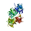
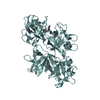
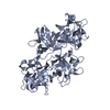
| Title | CRYSTAL STRUCTURE OF HUMAN FASCIN, AN ACTIN-CROSSLINKING PROTEIN | ||||||
 Components Components | FASCIN | ||||||
 Keywords Keywords | STRUCTURAL PROTEIN / BETA-TREFOIL FOLD FOR ALL FOUR DOMAINS / Structural Genomics / PSI / Protein Structure Initiative / New York SGX Research Center for Structural Genomics / NYSGXRC | ||||||
| Function / homology |  Function and homology information Function and homology informationmicrospike / parallel actin filament bundle assembly / regulation of microvillus assembly / microspike assembly / establishment of apical/basal cell polarity / positive regulation of extracellular matrix disassembly / cell projection membrane / positive regulation of podosome assembly / cell-cell junction assembly / podosome ...microspike / parallel actin filament bundle assembly / regulation of microvillus assembly / microspike assembly / establishment of apical/basal cell polarity / positive regulation of extracellular matrix disassembly / cell projection membrane / positive regulation of podosome assembly / cell-cell junction assembly / podosome / positive regulation of filopodium assembly / microvillus / establishment or maintenance of cell polarity / actin filament bundle assembly / positive regulation of lamellipodium assembly / ruffle / stress fiber / regulation of actin cytoskeleton organization / cell motility / filopodium / cell-cell junction / actin filament binding / cell migration / lamellipodium / actin cytoskeleton / growth cone / actin cytoskeleton organization / actin binding / Interleukin-4 and Interleukin-13 signaling / cell cortex / cytoskeleton / protein-macromolecule adaptor activity / cadherin binding / RNA binding / extracellular exosome / cytosol / cytoplasm Similarity search - Function | ||||||
| Biological species |  Homo sapiens (human) Homo sapiens (human) | ||||||
| Method |  X-RAY DIFFRACTION / MIR / Resolution: 2.9 Å X-RAY DIFFRACTION / MIR / Resolution: 2.9 Å | ||||||
 Authors Authors | Fedorov, A.A. / Fedorov, E.V. / Ono, S. / Matsumura, F. / Almo, S.C. / Burley, S.K. / New York SGX Research Center for Structural Genomics (NYSGXRC) | ||||||
 Citation Citation | Journal: J.Mol.Biol. / Year: 2010 Title: Structure, evolutionary conservation, and conformational dynamics of Homo sapiens fascin-1, an F-actin crosslinking protein. Authors: Sedeh, R.S. / Fedorov, A.A. / Fedorov, E.V. / Ono, S. / Matsumura, F. / Almo, S.C. / Bathe, M. #1: Journal: J.Biol.Chem. / Year: 1997Title: Identification of an Actin Binding Region and a Protein Kinase C Phosphorylation Site on Human Fascin Authors: Ono, S. / Yamakita, Y. / Yamashiro, S. / Matsudaira, P.T. / Gnarra, J.R. / Obinata, T. / Matsumura, F. | ||||||
| History |
|
- Structure visualization
| Structure viewer | Molecule: MolmilJmol/JSmol |
|---|
- Downloads & links
-Download
| PDBx/mmCIF format | 1dfc.cif.gz | 193.3 KB | Display | PDBx/mmCIF format |
|---|---|---|---|---|
| PDB format | pdb1dfc.ent.gz | 155.2 KB | Display | PDB format |
| PDBx/mmJSON format | 1dfc.json.gz | Tree view | PDBx/mmJSON format | |
| Others |  Other downloads Other downloads |
-Validation report
| Arichive directory | https://data.pdbj.org/pub/pdb/validation_reports/df/1dfcftp://data.pdbj.org/pub/pdb/validation_reports/df/1dfc | HTTPS FTP |
|---|
-Related structure data
| Similar structure data | |
|---|---|
| Other databases |


-Links
 PDBj
PDBj- Assembly
| Deposited unit | 
| ||||||||
|---|---|---|---|---|---|---|---|---|---|
| 1 | 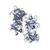
| ||||||||
| 2 | 
| ||||||||
| Unit cell |
|
-Components
| #1: Protein | Mass: 54601.879 Da / Num. of mol.: 2 / Fragment: FULL-LENGTH PROTEIN Source method: isolated from a genetically manipulated source Source: (gene. exp.) Homo sapiens (human) / Plasmid: PGEX-2T / Production host:  |
|---|
-Experimental details
-Experiment
| Experiment | Method: X-RAY DIFFRACTION / Number of used crystals: 1 |
|---|
- Sample preparation
| Crystal | Density Matthews: 2.3 Å3/Da / Density % sol: 46.6 % | ||||||||||||||||||||||||||||||||||||||||||||||||||||||||||||||||||||||
|---|---|---|---|---|---|---|---|---|---|---|---|---|---|---|---|---|---|---|---|---|---|---|---|---|---|---|---|---|---|---|---|---|---|---|---|---|---|---|---|---|---|---|---|---|---|---|---|---|---|---|---|---|---|---|---|---|---|---|---|---|---|---|---|---|---|---|---|---|---|---|---|
| Crystal grow | Temperature: 298 K / Method: vapor diffusion, hanging drop / pH: 7.5 Details: PEG 4000, hepes, dtt, sodium azide , pH 7.5, VAPOR DIFFUSION, HANGING DROP, temperature 298.0K | ||||||||||||||||||||||||||||||||||||||||||||||||||||||||||||||||||||||
| Crystal grow | *PLUS pH: 8 / Method: vapor diffusion, hanging drop | ||||||||||||||||||||||||||||||||||||||||||||||||||||||||||||||||||||||
| Components of the solutions | *PLUS
|
-Data collection
| Diffraction | Mean temperature: 291 K |
|---|---|
| Diffraction source | Source: ROTATING ANODE / Type: RIGAKU RU200 / Wavelength: 1.5418 |
| Detector | Type: SIEMENS / Detector: AREA DETECTOR / Date: Jun 19, 1997 |
| Radiation | Protocol: SINGLE WAVELENGTH / Monochromatic (M) / Laue (L): M / Scattering type: x-ray |
| Radiation wavelength | Wavelength: 1.5418 Å / Relative weight: 1 |
| Reflection | Resolution: 2.9→23.03 Å / Num. obs: 19147 / % possible obs: 84.2 % / Observed criterion σ(I): 1 / Redundancy: 2.6 % / Biso Wilson estimate: 22.8 Å2 / Rmerge(I) obs: 0.062 / Net I/σ(I): 19.1 |
| Reflection | *PLUS Highest resolution: 2.9 Å / Num. obs: 21421 / % possible obs: 94.2 % / Rmerge(I) obs: 0.048 |
- Processing
| Software |
| ||||||||||||||||||||||||||||||||||||||||||||||||||||||||||||||||||||||||||||||||
|---|---|---|---|---|---|---|---|---|---|---|---|---|---|---|---|---|---|---|---|---|---|---|---|---|---|---|---|---|---|---|---|---|---|---|---|---|---|---|---|---|---|---|---|---|---|---|---|---|---|---|---|---|---|---|---|---|---|---|---|---|---|---|---|---|---|---|---|---|---|---|---|---|---|---|---|---|---|---|---|---|---|
| Refinement | Method to determine structure: MIR / Resolution: 2.9→8 Å / σ(F): 0 / Stereochemistry target values: ENGH & HUBER
| ||||||||||||||||||||||||||||||||||||||||||||||||||||||||||||||||||||||||||||||||
| Solvent computation | Bsol: 63.17 Å2 / ksol: 0.247 e/Å3 | ||||||||||||||||||||||||||||||||||||||||||||||||||||||||||||||||||||||||||||||||
| Refine analyze | Luzzati coordinate error free: 0.343 Å | ||||||||||||||||||||||||||||||||||||||||||||||||||||||||||||||||||||||||||||||||
| Refinement step | Cycle: LAST / Resolution: 2.9→8 Å
| ||||||||||||||||||||||||||||||||||||||||||||||||||||||||||||||||||||||||||||||||
| Refine LS restraints |
| ||||||||||||||||||||||||||||||||||||||||||||||||||||||||||||||||||||||||||||||||
| Xplor file | Serial no: 1 / Param file: PROTEIN_REP.PARAM / Topol file: PROTEIN.TOP | ||||||||||||||||||||||||||||||||||||||||||||||||||||||||||||||||||||||||||||||||
| Refinement | *PLUS Highest resolution: 2.9 Å / Lowest resolution: 25 Å / Num. reflection obs: 20541 / Rfactor obs: 0.184 / Rfactor Rfree: 0.268 / Rfactor Rwork: 0.223 | ||||||||||||||||||||||||||||||||||||||||||||||||||||||||||||||||||||||||||||||||
| Solvent computation | *PLUS | ||||||||||||||||||||||||||||||||||||||||||||||||||||||||||||||||||||||||||||||||
| Displacement parameters | *PLUS | ||||||||||||||||||||||||||||||||||||||||||||||||||||||||||||||||||||||||||||||||
| Refine LS restraints | *PLUS
|
