 Movie
Movie Controller


+ Open data

- Basic information
| Entry | Database: PDB / ID: 1dcd | ||||||
|---|---|---|---|---|---|---|---|
| Title | DESULFOREDOXIN COMPLEXED WITH CD2+ | ||||||
 Components Components | PROTEIN (DESULFOREDOXIN) | ||||||
 Keywords Keywords | ELECTRON TRANSPORT / RUBREDOXIN TYPE PROTEIN | ||||||
| Function / homology | Desulforedoxin / Desulfoferrodoxin, N-terminal domain / Desulfoferrodoxin, N-terminal domain superfamily / Desulfoferrodoxin, N-terminal domain / iron ion binding / : / Desulforedoxin Function and homology information Function and homology information | ||||||
| Biological species |  Desulfovibrio gigas (bacteria) Desulfovibrio gigas (bacteria) | ||||||
| Method |  X-RAY DIFFRACTION / MOLECULAR REPLACEMENT / Resolution: 2 Å X-RAY DIFFRACTION / MOLECULAR REPLACEMENT / Resolution: 2 Å | ||||||
 Authors Authors | Archer, M. / Carvalho, A.L. / Teixeira, S. / Romao, M.J. | ||||||
 Citation Citation | Journal: Protein Sci. / Year: 1999 Title: Structural studies by X-ray diffraction on metal substituted desulforedoxin, a rubredoxin-type protein. Authors: Archer, M. / Carvalho, A.L. / Teixeira, S. / Moura, I. / Moura, J.J. / Rusnak, F. / Romao, M.J. #1: Journal: J.Mol.Biol. / Year: 1995Title: Crystal structure of desulforedoxin from Desulfovibrio gigas determined at 1.8 A resolution: a novel non-heme iron protein structure. Authors: Archer, M. / Huber, R. / Tavares, P. / Moura, I. / Moura, J.J. / Carrondo, M.A. / Sieker, L.C. / LeGall, J. / Romao, M.J. | ||||||
| History |
|
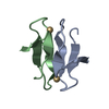
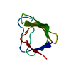
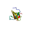
- Structure visualization
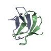
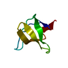
| Structure viewer | Molecule: MolmilJmol/JSmol |
|---|
- Downloads & links
-Download
| PDBx/mmCIF format | 1dcd.cif.gz | 25.5 KB | Display | PDBx/mmCIF format |
|---|---|---|---|---|
| PDB format | pdb1dcd.ent.gz | 16.5 KB | Display | PDB format |
| PDBx/mmJSON format | 1dcd.json.gz | Tree view | PDBx/mmJSON format | |
| Others |  Other downloads Other downloads |
-Validation report
| Arichive directory | https://data.pdbj.org/pub/pdb/validation_reports/dc/1dcdftp://data.pdbj.org/pub/pdb/validation_reports/dc/1dcd | HTTPS FTP |
|---|
-Related structure data
| Related structure data | 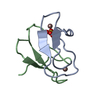 1cfwC  1dhgC 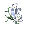 1dxgS S: Starting model for refinement C: citing same article ( |
|---|---|
| Similar structure data |


-Links
 PDBj
PDBj- Assembly
| Deposited unit | 
| ||||||||||
|---|---|---|---|---|---|---|---|---|---|---|---|
| 1 |
| ||||||||||
| Unit cell |
| ||||||||||
| Components on special symmetry positions |
| ||||||||||
| Noncrystallographic symmetry (NCS) | NCS oper: (Code: given Matrix: (-0.939027, -0.226955, -0.258303), Vector: |
-Components
| #1: Protein/peptide | Mass: 3807.350 Da / Num. of mol.: 2 Source method: isolated from a genetically manipulated source Source: (gene. exp.) Desulfovibrio gigas (bacteria) / Cellular location: CYTOPLASM / Cellular location (production host): CYTOPLASM / Gene (production host): DSR / Production host: #2: Chemical |   Mass: 112.411 Da / Num. of mol.: 2 / Source method: obtained synthetically / Formula: Cd Mass: 112.411 Da / Num. of mol.: 2 / Source method: obtained synthetically / Formula: Cd#3: Water | ChemComp-HOH / |  Mass: 18.015 Da / Num. of mol.: 53 / Source method: isolated from a natural source / Formula: H2O Mass: 18.015 Da / Num. of mol.: 53 / Source method: isolated from a natural source / Formula: H2O |
|---|
-Experimental details
-Experiment
| Experiment | Method: X-RAY DIFFRACTION / Number of used crystals: 1 |
|---|
- Sample preparation
| Crystal | Density Matthews: 1.9 Å3/Da / Density % sol: 35 % | ||||||||||||||||||||||||||||||||||||
|---|---|---|---|---|---|---|---|---|---|---|---|---|---|---|---|---|---|---|---|---|---|---|---|---|---|---|---|---|---|---|---|---|---|---|---|---|---|
| Crystal grow | pH: 5 Details: 30% ETHANOL, 0.1 M SODIUM ACETATE PH 5, 0.2 M CACL2, pH 5.0 | ||||||||||||||||||||||||||||||||||||
| Crystal grow | *PLUS pH: 7.6 / Method: vapor diffusion | ||||||||||||||||||||||||||||||||||||
| Components of the solutions | *PLUS
|
-Data collection
| Diffraction | Mean temperature: 277 K |
|---|---|
| Diffraction source | Source: ROTATING ANODE / Type: RIGAKU / Wavelength: 1.5418 |
| Detector | Type: MARRESEARCH / Detector: IMAGE PLATE |
| Radiation | Monochromator: NI FILTER / Protocol: SINGLE WAVELENGTH / Monochromatic (M) / Laue (L): M / Scattering type: x-ray |
| Radiation wavelength | Wavelength: 1.5418 Å / Relative weight: 1 |
| Reflection | Resolution: 1.98→14.8 Å / Num. all: 4403 / Num. obs: 4403 / % possible obs: 97.5 % / Observed criterion σ(I): 0 / Redundancy: 6.4 % / Rsym value: 0.115 / Net I/σ(I): 8 |
| Reflection shell | Resolution: 1.98→2.05 Å / Mean I/σ(I) obs: 2.5 / Rsym value: 0.432 / % possible all: 96.4 |
| Reflection | *PLUS Num. measured all: 28142 / Rmerge(I) obs: 0.115 |
| Reflection shell | *PLUS % possible obs: 96.4 % / Rmerge(I) obs: 0.359 |
- Processing
| Software |
| |||||||||||||||||||||||||||||||||
|---|---|---|---|---|---|---|---|---|---|---|---|---|---|---|---|---|---|---|---|---|---|---|---|---|---|---|---|---|---|---|---|---|---|---|
| Refinement | Method to determine structure: MOLECULAR REPLACEMENT Starting model: PDB ENTRY 1DXG Resolution: 2→14.8 Å / Num. parameters: 2325 / Num. restraintsaints: 2656 / σ(F): 0 / Stereochemistry target values: ENGH & HUBER Details: GEOMETRIC RESTRAINTS WERE APPLIED TO THE METAL CENTER (DFIX CD S 2.55 A)
| |||||||||||||||||||||||||||||||||
| Solvent computation | Solvent model: MOEWS & KRETSINGER, J.MOL.BIOL.91(1973)201-228 | |||||||||||||||||||||||||||||||||
| Refine analyze | Num. disordered residues: 2 / Occupancy sum hydrogen: 0 / Occupancy sum non hydrogen: 576 | |||||||||||||||||||||||||||||||||
| Refinement step | Cycle: LAST / Resolution: 2→14.8 Å
| |||||||||||||||||||||||||||||||||
| Refine LS restraints |
|
