 Movie
Movie Controller

[English] 日本語
 Yorodumi
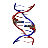
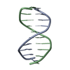
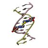
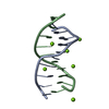
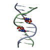
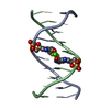
- PDB-1d62: THE STRUCTURE OF A /B-DNA$ DECAMER WITH AN I(SLASH)*A MISMATCH AN... -
+ Open data

- Basic information
| Entry | Database: PDB / ID: 1d62 | |||||||||||||||||||||
|---|---|---|---|---|---|---|---|---|---|---|---|---|---|---|---|---|---|---|---|---|---|---|
| Title | THE STRUCTURE OF A /B-DNA$ DECAMER WITH AN I(SLASH)*A MISMATCH AND COMPARISON WITH THE G(SLASH)*A MISMATCH | |||||||||||||||||||||
 Components Components | 5'-D(* Keywords KeywordsDNA | Function / homology | DNA |  Function and homology information Function and homology informationMethod |  X-RAY DIFFRACTION / Resolution: 2 Å X-RAY DIFFRACTION / Resolution: 2 Å  Authors AuthorsLipanov, A. / Kopka, M.L. / Kaczor-Grzeskowiak, M. / Dickerson, R.E. |  CitationJournal: Biochemistry / Year: 1993 CitationJournal: Biochemistry / Year: 1993Title: Structure of the B-DNA decamer C-C-A-A-C-I-T-T-G-G in two different space groups: conformational flexibility of B-DNA. Authors: Lipanov, A.A. / Kopka, M.L. / Kaczor-Grzeskowiak, M. / Dickerson, R.E. #1: Journal: J.Mol.Biol. / Year: 1991Title: The Structure of the /B-DNA Decamer / C-C-A-A-C-G-T-T-G-G and Comparison with the Isomorphous Decamers /C-C-A-A-G-A-T-T-G-G and /C-C-A-G-G-C-C-T-G-G Authors: Prive, G.G. / Yanagi, K. / Dickerson, R.E. #2: Journal: J.Mol.Biol. / Year: 1989Title: Crystallographic Study of One Turn of G(Slash)C-Rich B-/DNA Authors: Heinemann, U. / Alings, C. #3: Journal: Science / Year: 1987Title: Helix Geometry, Hydration, and G(Dot)A Mismatch in a B-/DNA Decamer Authors: Prive, G.G. / Heinemann, U. / Chandrasegaran, S. / Kan, L.-S. / Kopka, M.L. / Dickerson, R.E. History |
|
- Structure visualization
| Structure viewer | Molecule: MolmilJmol/JSmol |
|---|
- Downloads & links
-Download
| PDBx/mmCIF format | 1d62.cif.gz | 13.7 KB | Display | PDBx/mmCIF format |
|---|---|---|---|---|
| PDB format | pdb1d62.ent.gz | 9.2 KB | Display | PDB format |
| PDBx/mmJSON format | 1d62.json.gz | Tree view | PDBx/mmJSON format | |
| Others |  Other downloads Other downloads |
-Validation report
| Arichive directory | https://data.pdbj.org/pub/pdb/validation_reports/d6/1d62ftp://data.pdbj.org/pub/pdb/validation_reports/d6/1d62 | HTTPS FTP |
|---|
-Related structure data


-Links
 PDBj
PDBj

- Assembly
| Deposited unit | 
| |||||||||
|---|---|---|---|---|---|---|---|---|---|---|
| 1 | 
| |||||||||
| Unit cell |
| |||||||||
| Components on special symmetry positions |
|
-Components
| #1: DNA chain | Mass: 3054.016 Da / Num. of mol.: 1 / Source method: obtained synthetically |
|---|---|
| #2: Water | ChemComp-HOH /  Mass: 18.015 Da / Num. of mol.: 50 / Source method: isolated from a natural source / Formula: H2O Mass: 18.015 Da / Num. of mol.: 50 / Source method: isolated from a natural source / Formula: H2O |
-Experimental details
-Experiment
| Experiment | Method: X-RAY DIFFRACTION |
|---|
- Sample preparation
| Crystal | Density Matthews: 1.97 Å3/Da / Density % sol: 37.67 % | ||||||||||||||||||||||||||||||||||||||||||
|---|---|---|---|---|---|---|---|---|---|---|---|---|---|---|---|---|---|---|---|---|---|---|---|---|---|---|---|---|---|---|---|---|---|---|---|---|---|---|---|---|---|---|---|
| Crystal grow | *PLUS Temperature: 4 ℃ / Method: vapor diffusion, sitting drop / pH: 7 | ||||||||||||||||||||||||||||||||||||||||||
| Components of the solutions | *PLUS
|
-Data collection
| Radiation | Protocol: SINGLE WAVELENGTH / Monochromatic (M) / Laue (L): M / Scattering type: x-ray |
|---|---|
| Radiation wavelength | Relative weight: 1 |
| Reflection | *PLUS |
- Processing
| Software | Name: NUCLSQ / Classification: refinement | ||||||||||||||||||||
|---|---|---|---|---|---|---|---|---|---|---|---|---|---|---|---|---|---|---|---|---|---|
| Refinement | Resolution: 2→8 Å / σ(F): 2 /
| ||||||||||||||||||||
| Refinement step | Cycle: LAST / Resolution: 2→8 Å
| ||||||||||||||||||||
| Software | *PLUS Name: NUCLSQ / Classification: refinement | ||||||||||||||||||||
| Refinement | *PLUS Highest resolution: 2 Å / Num. reflection obs: 1195 / Rfactor obs: 0.134 | ||||||||||||||||||||
| Solvent computation | *PLUS | ||||||||||||||||||||
| Displacement parameters | *PLUS | ||||||||||||||||||||
| Refine LS restraints | *PLUS
|
