 Movie
Movie Controller

[English] 日本語
 Yorodumi
- PDB-1d1w: BOVINE ENDOTHELIAL NITRIC OXIDE SYNTHASE HEME DOMAIN COMPLEXED WI... -
+ Open data

- Basic information
| Entry | Database: PDB / ID: 1d1w | ||||||
|---|---|---|---|---|---|---|---|
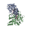
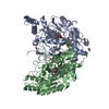
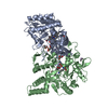
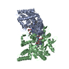
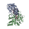
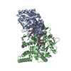
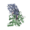
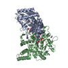
| Title | BOVINE ENDOTHELIAL NITRIC OXIDE SYNTHASE HEME DOMAIN COMPLEXED WITH 2-AMINOTHIAZOLINE (H4B BOUND) | ||||||
 Components Components | NITRIC OXIDE SYNTHASE | ||||||
 Keywords Keywords | OXIDOREDUCTASE / ALPHA-BETA FOLD | ||||||
| Function / homology |  Function and homology information Function and homology informationcellular response to laminar fluid shear stress / negative regulation of leukocyte cell-cell adhesion / nitric-oxide synthase (NADPH) / : / nitric-oxide synthase activity / L-arginine catabolic process / negative regulation of extrinsic apoptotic signaling pathway via death domain receptors / nitric oxide biosynthetic process / negative regulation of blood pressure / response to hormone ...cellular response to laminar fluid shear stress / negative regulation of leukocyte cell-cell adhesion / nitric-oxide synthase (NADPH) / : / nitric-oxide synthase activity / L-arginine catabolic process / negative regulation of extrinsic apoptotic signaling pathway via death domain receptors / nitric oxide biosynthetic process / negative regulation of blood pressure / response to hormone / positive regulation of relaxation of smooth muscle / mitochondrion organization / caveola / response to peptide hormone / blood coagulation / NADP binding / FMN binding / flavin adenine dinucleotide binding / response to lipopolysaccharide / cytoskeleton / calmodulin binding / heme binding / Golgi apparatus / metal ion binding / nucleus / plasma membrane / cytosol Similarity search - Function | ||||||
| Biological species |  | ||||||
| Method |  X-RAY DIFFRACTION / SYNCHROTRON / Resolution: 2 Å X-RAY DIFFRACTION / SYNCHROTRON / Resolution: 2 Å | ||||||
 Authors Authors | Li, H. / Raman, C.S. / Martasek, P. / Kral, V. / Masters, B.S.S. / Poulos, T.L. | ||||||
 Citation Citation | Journal: J.Inorg.Biochem. / Year: 2000 Title: Mapping the active site polarity in structures of endothelial nitric oxide synthase heme domain complexed with isothioureas. Authors: Li, H. / Raman, C.S. / Martasek, P. / Kral, V. / Masters, B.S. / Poulos, T.L. #1: Journal: Cell(Cambridge,Mass.) / Year: 1998Title: Crystal Structure of Constitutive Endothelial Nitric Oxide Synthase: a Paradigm for Pterin Function Involving a Novel Metal Center Authors: Raman, C.S. / Li, H. / Martasek, P. / Kral, V. / Masters, B.S. / Poulos, T.L. #2: Journal: Science / Year: 1998Title: Structure of Nitric Oxide Synthase Oxygenase Dimer with Pterin and Substrate Authors: Crane, B.R. / Arvai, A.S. / Ghosh, D.K. / Wu, C. / Getzoff, E.D. / Stuehr, D.J. / Tainer, J.A. #3: Journal: Br.J.Pharmacol. / Year: 1996Title: Spontaneous Rearrangement of Aminoalkylisothioureas into Mercaptoalkylguanidines, a Novel Class of Nitric Oxide Synthase Inhibitors with Selectivity Towards the Inducible Isoform Authors: Southan, G.J. / Zingarelli, B. / O'Connor, M. / Salzman, A.L. / Szabo, C. | ||||||
| History |
|
- Structure visualization
| Structure viewer | Molecule: MolmilJmol/JSmol |
|---|
- Downloads & links
-Download
| PDBx/mmCIF format | 1d1w.cif.gz | 193 KB | Display | PDBx/mmCIF format |
|---|---|---|---|---|
| PDB format | pdb1d1w.ent.gz | 151.1 KB | Display | PDB format |
| PDBx/mmJSON format | 1d1w.json.gz | Tree view | PDBx/mmJSON format | |
| Others |  Other downloads Other downloads |
-Validation report
| Arichive directory | https://data.pdbj.org/pub/pdb/validation_reports/d1/1d1wftp://data.pdbj.org/pub/pdb/validation_reports/d1/1d1w | HTTPS FTP |
|---|
-Related structure data


-Links
 PDBj
PDBj


- Assembly
| Deposited unit | 
| ||||||||
|---|---|---|---|---|---|---|---|---|---|
| 1 |
| ||||||||
| Unit cell |
|
-Components
-Protein , 1 types, 2 molecules AB
| #1: Protein | Mass: 49710.105 Da / Num. of mol.: 2 / Fragment: HEME DOMAIN Source method: isolated from a genetically manipulated source Source: (gene. exp.)  |
|---|
-Non-polymers , 8 types, 433 molecules 


| #2: Chemical | ChemComp-ACT /  Mass: 59.044 Da / Num. of mol.: 4 / Source method: obtained synthetically / Formula: C2H3O2 Mass: 59.044 Da / Num. of mol.: 4 / Source method: obtained synthetically / Formula: C2H3O2#3: Chemical | ChemComp-ZN / |  Mass: 65.409 Da / Num. of mol.: 1 / Source method: obtained synthetically / Formula: Zn Mass: 65.409 Da / Num. of mol.: 1 / Source method: obtained synthetically / Formula: Zn#4: Chemical |  Mass: 616.487 Da / Num. of mol.: 2 / Source method: obtained synthetically / Formula: C34H32FeN4O4 Mass: 616.487 Da / Num. of mol.: 2 / Source method: obtained synthetically / Formula: C34H32FeN4O4#5: Chemical |  Mass: 241.247 Da / Num. of mol.: 2 / Source method: obtained synthetically / Formula: C9H15N5O3 Mass: 241.247 Da / Num. of mol.: 2 / Source method: obtained synthetically / Formula: C9H15N5O3#6: Chemical |  Mass: 102.158 Da / Num. of mol.: 2 / Source method: obtained synthetically / Formula: C3H6N2S Mass: 102.158 Da / Num. of mol.: 2 / Source method: obtained synthetically / Formula: C3H6N2S#7: Chemical |  Mass: 137.997 Da / Num. of mol.: 2 / Source method: obtained synthetically / Formula: C2H7AsO2 Mass: 137.997 Da / Num. of mol.: 2 / Source method: obtained synthetically / Formula: C2H7AsO2#8: Chemical | ChemComp-GOL /  Mass: 92.094 Da / Num. of mol.: 4 / Source method: obtained synthetically / Formula: C3H8O3 Mass: 92.094 Da / Num. of mol.: 4 / Source method: obtained synthetically / Formula: C3H8O3#9: Water | ChemComp-HOH / | Mass: 18.015 Da / Num. of mol.: 416 / Source method: isolated from a natural source / Formula: H2O |
|---|
-Experimental details
-Experiment
| Experiment | Method: X-RAY DIFFRACTION / Number of used crystals: 1 |
|---|
- Sample preparation
| Crystal | Density Matthews: 2.44 Å3/Da / Density % sol: 49.62 % | ||||||||||||||||||||||||||||||||||||||||||
|---|---|---|---|---|---|---|---|---|---|---|---|---|---|---|---|---|---|---|---|---|---|---|---|---|---|---|---|---|---|---|---|---|---|---|---|---|---|---|---|---|---|---|---|
| Crystal grow | Temperature: 280 K / Method: vapor diffusion, sitting drop / pH: 6.5 Details: PEG 4000, CACODYLATE, MAGNESIUM ACETATE THE CHEMICAL, S-AMINOETHYL-ISOTHIOUREA, ADDED TO THE CRYSTALLIZATION BUFFER UNDERGOES A SPONTANEOUS REARRANGEMENT TO FORM 2-AMINOTHIAZOLINE., pH 6.5, ...Details: PEG 4000, CACODYLATE, MAGNESIUM ACETATE THE CHEMICAL, S-AMINOETHYL-ISOTHIOUREA, ADDED TO THE CRYSTALLIZATION BUFFER UNDERGOES A SPONTANEOUS REARRANGEMENT TO FORM 2-AMINOTHIAZOLINE., pH 6.5, VAPOR DIFFUSION, SITTING DROP, temperature 280K | ||||||||||||||||||||||||||||||||||||||||||
| Crystal grow | *PLUS | ||||||||||||||||||||||||||||||||||||||||||
| Components of the solutions | *PLUS
|
-Data collection
| Diffraction | Mean temperature: 100 K |
|---|---|
| Diffraction source | Source: SYNCHROTRON / Site: SSRL  / Beamline: BL7-1 / Wavelength: 1.08 / Beamline: BL7-1 / Wavelength: 1.08 |
| Detector | Type: MARRESEARCH / Detector: IMAGE PLATE / Date: Apr 13, 1999 |
| Radiation | Protocol: SINGLE WAVELENGTH / Monochromatic (M) / Laue (L): M / Scattering type: x-ray |
| Radiation wavelength | Wavelength: 1.08 Å / Relative weight: 1 |
| Reflection | Resolution: 2→50 Å / Num. all: 66684 / Num. obs: 61326 / % possible obs: 89.7 % / Observed criterion σ(I): -3 / Redundancy: 2.8 % / Biso Wilson estimate: 25.6 Å2 / Rmerge(I) obs: 0.051 / Net I/σ(I): 9.3 |
| Reflection shell | Resolution: 2→2.03 Å / Redundancy: 2.2 % / Rmerge(I) obs: 0.7 / % possible all: 84.4 |
| Reflection | *PLUS Num. measured all: 174141 |
| Reflection shell | *PLUS % possible obs: 84.4 % |
- Processing
| Software |
| ||||||||||||||||||||||||||||||||||||||||||||||||||||||||||||
|---|---|---|---|---|---|---|---|---|---|---|---|---|---|---|---|---|---|---|---|---|---|---|---|---|---|---|---|---|---|---|---|---|---|---|---|---|---|---|---|---|---|---|---|---|---|---|---|---|---|---|---|---|---|---|---|---|---|---|---|---|---|
| Refinement | Resolution: 2→50 Å / Rfactor Rfree error: 0.005 / Cross valid method: THROUGHOUT / σ(F): 0 / Stereochemistry target values: ENGH & HUBER Details: RESIDUES 108-120 ARE DISORDERED, BUT THE TENTATIVE MODEL WAS INCLUDED IN THE REFINEMENT.
| ||||||||||||||||||||||||||||||||||||||||||||||||||||||||||||
| Solvent computation | Solvent model: flat model / Bsol: 56.9463 Å2 / ksol: 0.365778 e/Å3 | ||||||||||||||||||||||||||||||||||||||||||||||||||||||||||||
| Displacement parameters | Biso mean: 44.9 Å2
| ||||||||||||||||||||||||||||||||||||||||||||||||||||||||||||
| Refine analyze |
| ||||||||||||||||||||||||||||||||||||||||||||||||||||||||||||
| Refinement step | Cycle: LAST / Resolution: 2→50 Å
| ||||||||||||||||||||||||||||||||||||||||||||||||||||||||||||
| Refine LS restraints |
| ||||||||||||||||||||||||||||||||||||||||||||||||||||||||||||
| LS refinement shell | Resolution: 2→2.07 Å / Rfactor Rfree error: 0.027 / Total num. of bins used: 10
| ||||||||||||||||||||||||||||||||||||||||||||||||||||||||||||
| Software | *PLUS Name: CNS / Classification: refinement | ||||||||||||||||||||||||||||||||||||||||||||||||||||||||||||
| Refine LS restraints | *PLUS
|
