- PDB-1cyy: CRYSTAL STRUCTURE OF THE 30 KDA FRAGMENT OF E. COLI DNA TOPOISOME... -
+
Open data
ID or keywords:
Loading...
-
Basic information
Entry
Database: PDB / ID: 1cyy
Title
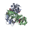
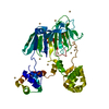
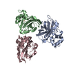
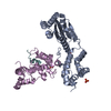
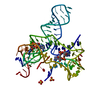
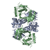
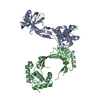
CRYSTAL STRUCTURE OF THE 30 KDA FRAGMENT OF E. COLI DNA TOPOISOMERASE I. HEXAGONAL FORM
Components
DNA TOPOISOMERASE I
Keywords
ISOMERASE / DNA TOPOISOMERASE / DECATENATING ENZYME
Function / homology
Function and homology information
RNA topoisomerase activity / DNA topoisomerase activity / DNA topoisomerase / DNA topoisomerase type I (single strand cut, ATP-independent) activity / DNA topological change / chromosome / DNA binding / zinc ion binding / cytosol Similarity search - Function
DNA topoisomerase I, zinc ribbon-like, bacterial-type / : / Topoisomerase I zinc-ribbon-like / Topoisomerase I, zinc finger / DNA topoisomerase, type IA, zn finger / Topoisomerase DNA binding C4 zinc finger / DNA topoisomerase I, bacterial-type / DNA topoisomerase I, type IA / DNA topoisomerase 1, TOPRIM domain / Topoisomerase I, domain 3 ...DNA topoisomerase I, zinc ribbon-like, bacterial-type / : / Topoisomerase I zinc-ribbon-like / Topoisomerase I, zinc finger / DNA topoisomerase, type IA, zn finger / Topoisomerase DNA binding C4 zinc finger / DNA topoisomerase I, bacterial-type / DNA topoisomerase I, type IA / DNA topoisomerase 1, TOPRIM domain / Topoisomerase I, domain 3 / Topoisomerase I; domain 4 / Topoisomerase I, domain 4 / Topoisomerase I; domain 3 / DNA topoisomerase, type IA / DNA topoisomerase, type IA, central region, subdomain 2 / DNA topoisomerase, type IA, active site / Topoisomerase (Topo) IA-type active site signature. / Topoisomerase (Topo) IA-type catalytic domain profile. / DNA topoisomerase, type IA, domain 2 / DNA topoisomerase, type IA, DNA-binding domain / DNA topoisomerase, type IA, central / DNA topoisomerase, type IA, central region, subdomain 1 / DNA topoisomerase, type IA, central region, subdomain 3 / DNA topoisomerase, type IA, core domain / DNA topoisomerase / Bacterial DNA topoisomeraes I ATP-binding domain / Bacterial DNA topoisomerase I DNA-binding domain / TOPRIM / Toprim domain / Toprim domain profile. / TOPRIM domain / Distorted Sandwich / Orthogonal Bundle / Mainly Beta / Mainly Alpha Similarity search - Domain/homology
Resolution: 2.15→2.21 Å / Redundancy: 3.8 % / Rmerge(I) obs: 0.261 / % possible all: 85.8
Reflection
*PLUS
Num. measured all: 259275
Reflection shell
*PLUS
% possible obs: 85.8 %
-
Processing
Software
Name
Version
Classification
CNS
0.9
refinement
REFMAC
refinement
X-PLOR
refinement
AMoRE
phasing
DENZO
datareduction
CCP4
(SCALA)
datascaling
Refinement
Resolution: 2.15→14.97 Å / Rfactor Rfree error: 0.006 / Data cutoff high absF: 330269769.3 / Data cutoff low absF: 0 / Isotropic thermal model: RESTRAINED / Cross valid method: THROUGHOUT / σ(F): 2 Stereochemistry target values: R. A. ENGH AND R. HUBER, ACTA CRYST. SECT. A., 1991 Details: MOST OF THE REFINEMENT DONE WITH REFMAC. SOLVENT CORRECTION WITH XPLOR. FINAL REFINEMENT WITH CNS 0.9
Rfactor
Num. reflection
% reflection
Selection details
Rfree
0.282
2166
5 %
RANDOM
Rwork
0.229
-
-
-
all
0.234
44947
-
-
obs
0.234
43571
88.3 %
-
Displacement parameters
Biso mean: 41.6 Å2
Baniso -1
Baniso -2
Baniso -3
1-
0.93 Å2
4.37 Å2
0 Å2
2-
-
0.93 Å2
0 Å2
3-
-
-
-1.86 Å2
Refine analyze
Free
Obs
Luzzati coordinate error
0.38 Å
0.29 Å
Luzzati d res low
-
5 Å
Luzzati sigma a
0.33 Å
0.24 Å
Refinement step
Cycle: LAST / Resolution: 2.15→14.97 Å
Protein
Nucleic acid
Ligand
Solvent
Total
Num. atoms
3939
0
1
380
4320
Refine LS restraints
Refine-ID
Type
Dev ideal
Dev ideal target
X-RAY DIFFRACTION
c_bond_d
0.008
X-RAY DIFFRACTION
c_bond_d_na
X-RAY DIFFRACTION
c_bond_d_prot
X-RAY DIFFRACTION
c_angle_d
X-RAY DIFFRACTION
c_angle_d_na
X-RAY DIFFRACTION
c_angle_d_prot
X-RAY DIFFRACTION
c_angle_deg
1.4
X-RAY DIFFRACTION
c_angle_deg_na
X-RAY DIFFRACTION
c_angle_deg_prot
X-RAY DIFFRACTION
c_dihedral_angle_d
23.7
X-RAY DIFFRACTION
c_dihedral_angle_d_na
X-RAY DIFFRACTION
c_dihedral_angle_d_prot
X-RAY DIFFRACTION
c_improper_angle_d
0.88
X-RAY DIFFRACTION
c_improper_angle_d_na
X-RAY DIFFRACTION
c_improper_angle_d_prot
X-RAY DIFFRACTION
c_mcbond_it
1.09
2
X-RAY DIFFRACTION
c_mcangle_it
1.89
3
X-RAY DIFFRACTION
c_scbond_it
1.16
2
X-RAY DIFFRACTION
c_scangle_it
2
3
LS refinement shell
Resolution: 2.15→2.23 Å / Rfactor Rfree error: 0.029 / Total num. of bins used: 10
Rfactor
Num. reflection
% reflection
Rfree
0.382
178
4.6 %
Rwork
0.288
3732
-
obs
-
-
81.2 %
Xplor file
Refine-ID
Serial no
Param file
Topol file
X-RAY DIFFRACTION
1
PROTEIN_REP.PA
PROTEIN.TOP
X-RAY DIFFRACTION
2
WATER_REP.PARA
WATER.TOP
X-RAY DIFFRACTION
3
ION.PARAM
ION.TOP
Software
*PLUS
Name: 'REFMAC, XPLOR AND CNS 0.9' / Classification: refinement
Refine LS restraints
*PLUS
Refine-ID
Type
Dev ideal
X-RAY DIFFRACTION
p_bond_d
X-RAY DIFFRACTION
p_angle_d
X-RAY DIFFRACTION
p_angle_deg
X-RAY DIFFRACTION
p_dihedral_angle_d
X-RAY DIFFRACTION
p_dihedral_angle_deg
23.7
X-RAY DIFFRACTION
p_improper_angle_d
X-RAY DIFFRACTION
p_improper_angle_deg
0.88
X-RAY DIFFRACTION
p_mcbond_it
X-RAY DIFFRACTION
p_scbond_it
X-RAY DIFFRACTION
p_mcangle_it
X-RAY DIFFRACTION
p_scangle_it
+
About Yorodumi
-
News
-
Feb 9, 2022. New format data for meta-information of EMDB entries
New format data for meta-information of EMDB entries
Version 3 of the EMDB header file is now the official format.
The previous official version 1.9 will be removed from the archive.
In the structure databanks used in Yorodumi, some data are registered as the other names, "COVID-19 virus" and "2019-nCoV". Here are the details of the virus and the list of structure data.
Jan 31, 2019. EMDB accession codes are about to change! (news from PDBe EMDB page)
EMDB accession codes are about to change! (news from PDBe EMDB page)
The allocation of 4 digits for EMDB accession codes will soon come to an end. Whilst these codes will remain in use, new EMDB accession codes will include an additional digit and will expand incrementally as the available range of codes is exhausted. The current 4-digit format prefixed with “EMD-” (i.e. EMD-XXXX) will advance to a 5-digit format (i.e. EMD-XXXXX), and so on. It is currently estimated that the 4-digit codes will be depleted around Spring 2019, at which point the 5-digit format will come into force.
The EM Navigator/Yorodumi systems omit the EMD- prefix.
Related info.:Q: What is EMD? / ID/Accession-code notation in Yorodumi/EM Navigator
Yorodumi is a browser for structure data from EMDB, PDB, SASBDB, etc.
This page is also the successor to EM Navigator detail page, and also detail information page/front-end page for Omokage search.
The word "yorodu" (or yorozu) is an old Japanese word meaning "ten thousand". "mi" (miru) is to see.
Related info.:EMDB / PDB / SASBDB / Comparison of 3 databanks / Yorodumi Search / Aug 31, 2016. New EM Navigator & Yorodumi / Yorodumi Papers / Jmol/JSmol / Function and homology information / Changes in new EM Navigator and Yorodumi
 Movie
Movie Controller

 Yorodumi
 Open data
 Basic information
 Components
 Keywords
 Function and homology information
 X-RAY DIFFRACTION /
  Authors
 Citation
 Structure visualization
 Downloads & links
 Other downloads


 PDBj
PDBj


 Assembly
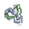

 Mass: 65.409 Da / Num. of mol.: 1 / Source method: obtained synthetically / Formula: Zn
 Mass: 18.015 Da / Num. of mol.: 380 / Source method: isolated from a natural source / Formula: H2O
 Sample preparation
 / Beamline: 5ID-B / Wavelength: 1.002
  Processing