 Movie
Movie Controller

[English] 日本語
 Yorodumi
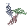
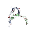
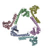
- PDB-1cun: CRYSTAL STRUCTURE OF REPEATS 16 AND 17 OF CHICKEN BRAIN ALPHA SPECTRIN -
+ Open data

- Basic information
| Entry | Database: PDB / ID: 1cun | ||||||
|---|---|---|---|---|---|---|---|
| Title | CRYSTAL STRUCTURE OF REPEATS 16 AND 17 OF CHICKEN BRAIN ALPHA SPECTRIN | ||||||
 Components Components | PROTEIN (ALPHA SPECTRIN) | ||||||
 Keywords Keywords | STRUCTURAL PROTEIN / TWO REPEATS OF SPECTRIN / ALPHA HELICAL LINKER REGION / 2 TANDEM 3-HELIX COILED- COILS | ||||||
| Function / homology |  Function and homology information Function and homology informationcostamere / actin filament capping / cortical actin cytoskeleton / cell projection / actin filament binding / cell junction / actin cytoskeleton organization / calmodulin binding / calcium ion binding / plasma membrane Similarity search - Function | ||||||
| Biological species |  | ||||||
| Method |  X-RAY DIFFRACTION / SYNCHROTRON / Resolution: 2 Å X-RAY DIFFRACTION / SYNCHROTRON / Resolution: 2 Å | ||||||
 Authors Authors | Grum, V.L. / Li, D. / MacDonald, R.I. / Mondragon, A. | ||||||
 Citation Citation | Journal: Cell(Cambridge,Mass.) / Year: 1999 Title: Structures of two repeats of spectrin suggest models of flexibility. Authors: Grum, V.L. / Li, D. / MacDonald, R.I. / Mondragon, A. | ||||||
| History |
|
- Structure visualization
| Structure viewer | Molecule: MolmilJmol/JSmol |
|---|
- Downloads & links
-Download
| PDBx/mmCIF format | 1cun.cif.gz | 148.6 KB | Display | PDBx/mmCIF format |
|---|---|---|---|---|
| PDB format | pdb1cun.ent.gz | 119 KB | Display | PDB format |
| PDBx/mmJSON format | 1cun.json.gz | Tree view | PDBx/mmJSON format | |
| Others |  Other downloads Other downloads |
-Validation report
| Arichive directory | https://data.pdbj.org/pub/pdb/validation_reports/cu/1cunftp://data.pdbj.org/pub/pdb/validation_reports/cu/1cun | HTTPS FTP |
|---|
-Related structure data
| Similar structure data |
|---|

-Links
 PDBj
PDBj- Assembly
| Deposited unit | 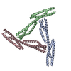
| ||||||||
|---|---|---|---|---|---|---|---|---|---|
| 1 |
| ||||||||
| Unit cell |
|
-Components
| #1: Protein | Mass: 24383.373 Da / Num. of mol.: 3 Fragment: REPEATS 16 AND 17 (RESIDUES 1771 TO 1982 PLUS AN N-TERMINAL MET) Mutation: L1771V, G1827S Source method: isolated from a genetically manipulated source Source: (gene. exp.)  #2: Water | ChemComp-HOH / |  Mass: 18.015 Da / Num. of mol.: 688 / Source method: isolated from a natural source / Formula: H2O Mass: 18.015 Da / Num. of mol.: 688 / Source method: isolated from a natural source / Formula: H2O |
|---|
-Experimental details
-Experiment
| Experiment | Method: X-RAY DIFFRACTION / Number of used crystals: 1 |
|---|
- Sample preparation
| Crystal | Density Matthews: 4.12 Å3/Da / Density % sol: 70.11 % | ||||||||||||||||||||||||||||||
|---|---|---|---|---|---|---|---|---|---|---|---|---|---|---|---|---|---|---|---|---|---|---|---|---|---|---|---|---|---|---|---|
| Crystal grow | Temperature: 277 K / Method: vapor diffusion, hanging drop / pH: 5.6 Details: 1.99M AMMONIUM SULFATE, 0.12M NA, K TARTRATE, 0.1M NA CITRATE PH 5.6, VAPOR DIFFUSION, HANGING DROP, temperature 277K | ||||||||||||||||||||||||||||||
| Crystal | *PLUS Density % sol: 70 % | ||||||||||||||||||||||||||||||
| Crystal grow | *PLUS Temperature: 4 ℃ / pH: 7.5 / Details: used microseeding | ||||||||||||||||||||||||||||||
| Components of the solutions | *PLUS
|
-Data collection
| Diffraction | Mean temperature: 108 K |
|---|---|
| Diffraction source | Source: SYNCHROTRON / Site: APS  / Beamline: 5ID-B / Wavelength: 1 / Beamline: 5ID-B / Wavelength: 1 |
| Detector | Type: MARRESEARCH / Detector: CCD / Date: May 1, 1998 |
| Radiation | Protocol: SINGLE WAVELENGTH / Monochromatic (M) / Laue (L): M / Scattering type: x-ray |
| Radiation wavelength | Wavelength: 1 Å / Relative weight: 1 |
| Reflection | Resolution: 1.95→30 Å / Num. obs: 387128 / % possible obs: 91.3 % / Observed criterion σ(I): 0 / Redundancy: 4.8 % / Biso Wilson estimate: 32.3 Å2 / Rmerge(I) obs: 0.055 / Net I/σ(I): 9.3 |
| Reflection shell | Resolution: 2→2.06 Å / Redundancy: 2.1 % / Rmerge(I) obs: 0.17 / % possible all: 79.5 |
| Reflection | *PLUS Highest resolution: 1.95 Å / Lowest resolution: 30 Å / Num. obs: 75300 / Observed criterion σ(I): 0 / Redundancy: 4.8 % / Num. measured all: 387128 |
| Reflection shell | *PLUS Redundancy: 2.1 % |
- Processing
| Software |
| ||||||||||||||||||||||||||||||||||||||||||||||||||||||||||||||||||||||||||||||||
|---|---|---|---|---|---|---|---|---|---|---|---|---|---|---|---|---|---|---|---|---|---|---|---|---|---|---|---|---|---|---|---|---|---|---|---|---|---|---|---|---|---|---|---|---|---|---|---|---|---|---|---|---|---|---|---|---|---|---|---|---|---|---|---|---|---|---|---|---|---|---|---|---|---|---|---|---|---|---|---|---|---|
| Refinement | Resolution: 2→24.63 Å / Rfactor Rfree error: 0.004 / Data cutoff high absF: 34928405.54 / Data cutoff low absF: 0 / Isotropic thermal model: RESTRAINED / Cross valid method: THROUGHOUT
| ||||||||||||||||||||||||||||||||||||||||||||||||||||||||||||||||||||||||||||||||
| Displacement parameters | Biso mean: 38.7 Å2
| ||||||||||||||||||||||||||||||||||||||||||||||||||||||||||||||||||||||||||||||||
| Refine analyze |
| ||||||||||||||||||||||||||||||||||||||||||||||||||||||||||||||||||||||||||||||||
| Refinement step | Cycle: LAST / Resolution: 2→24.63 Å
| ||||||||||||||||||||||||||||||||||||||||||||||||||||||||||||||||||||||||||||||||
| Refine LS restraints |
| ||||||||||||||||||||||||||||||||||||||||||||||||||||||||||||||||||||||||||||||||
| LS refinement shell | Resolution: 2→2.13 Å / Rfactor Rfree error: 0.013 / Total num. of bins used: 6
| ||||||||||||||||||||||||||||||||||||||||||||||||||||||||||||||||||||||||||||||||
| Xplor file |
| ||||||||||||||||||||||||||||||||||||||||||||||||||||||||||||||||||||||||||||||||
| Software | *PLUS Name: 'REFMAC, X-PLOR' / Classification: refinement | ||||||||||||||||||||||||||||||||||||||||||||||||||||||||||||||||||||||||||||||||
| Refinement | *PLUS % reflection Rfree: 5 % / Rfactor obs: 0.216 / Rfactor Rfree: 0.262 / Rfactor Rwork: 0.22 | ||||||||||||||||||||||||||||||||||||||||||||||||||||||||||||||||||||||||||||||||
| Solvent computation | *PLUS | ||||||||||||||||||||||||||||||||||||||||||||||||||||||||||||||||||||||||||||||||
| Displacement parameters | *PLUS Biso mean: 38.7 Å2 | ||||||||||||||||||||||||||||||||||||||||||||||||||||||||||||||||||||||||||||||||
| Refine LS restraints | *PLUS
|
