[English] 日本語
Yorodumi
- PDB-1crc: CYTOCHROME C AT LOW IONIC STRENGTH -

+
Open data


ID or keywords:

Loading...

-
Basic information

Entry
Database: PDB / ID: 1crc
TitleCYTOCHROME C AT LOW IONIC STRENGTH
ComponentsCYTOCHROME C
Keywords
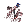
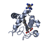
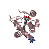
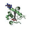
MITOCHONDRIAL ELECTRON TRANSPORT / FERRIC FORM / LOW IONIC STRENGTH
Function / homology
Function and homology information


cytochrome c-heme linkage / cytochrome complex / mitochondrial electron transport, cytochrome c to oxygen / mitochondrial electron transport, ubiquinol to cytochrome c / apoptotic signaling pathway / mitochondrial intermembrane space / electron transfer activity / lipid binding / heme binding / metal ion binding ...cytochrome c-heme linkage / cytochrome complex / mitochondrial electron transport, cytochrome c to oxygen / mitochondrial electron transport, ubiquinol to cytochrome c / apoptotic signaling pathway / mitochondrial intermembrane space / electron transfer activity / lipid binding / heme binding / metal ion binding / identical protein binding / cytosol
Similarity search - Function
Cytochrome c, class IA/ IB / Cytochrome c / Cytochrome c-like domain / Cytochrome Bc1 Complex; Chain D, domain 2 / Cytochrome c family profile. / Cytochrome c-like domain / Cytochrome c-like domain superfamily / Orthogonal Bundle / Mainly Alpha
Similarity search - Domain/homology
HEME C / Cytochrome c
Similarity search - Component
Biological speciesEquus caballus (horse)
MethodX-RAY DIFFRACTION / Resolution: 2.08 Å
AuthorsSanishvili, R. / Volz, K.W. / Westbrook, E.M. / Margoliash, E.
Citation
Journal: Structure / Year: 1995
Title: The low ionic strength crystal structure of horse cytochrome c at 2.1 A resolution and comparison with its high ionic strength counterpart.
Authors: Sanishvili, R. / Volz, K.W. / Westbrook, E.M. / Margoliash, E.
#1: Journal: Acta Crystallogr.,Sect.D / Year: 1994
Title: Crystallization of Wild-Type and Mutant Ferricytochromes C at Low Ionic Strength: Seeding Technique and X-Ray Diffraction Analysis
Authors: Sanishvili, R. / Margoliash, E. / Westbrook, M.L. / Westbrook, E.M. / Volz, K.W.
History
DepositionMar 22, 1995Processing site: BNL
Revision 1.0Mar 8, 1996Provider: repository / Type: Initial release
Revision 1.1Mar 24, 2008Group: Version format compliance
Revision 1.2Jul 13, 2011Group: Version format compliance
Revision 2.0Mar 10, 2021Group: Advisory / Atomic model ...Advisory / Atomic model / Data collection / Derived calculations / Non-polymer description / Other / Structure summary
Category: atom_site / chem_comp ...atom_site / chem_comp / entity / pdbx_database_status / pdbx_entity_nonpoly / pdbx_nonpoly_scheme / pdbx_struct_conn_angle / pdbx_validate_close_contact / struct_conn / struct_site
Item: _atom_site.B_iso_or_equiv / _atom_site.Cartn_x ..._atom_site.B_iso_or_equiv / _atom_site.Cartn_x / _atom_site.Cartn_y / _atom_site.Cartn_z / _atom_site.auth_atom_id / _atom_site.auth_comp_id / _atom_site.label_atom_id / _atom_site.label_comp_id / _atom_site.type_symbol / _chem_comp.formula / _chem_comp.formula_weight / _chem_comp.id / _chem_comp.name / _chem_comp.pdbx_synonyms / _entity.formula_weight / _entity.pdbx_description / _pdbx_database_status.process_site / _pdbx_entity_nonpoly.comp_id / _pdbx_entity_nonpoly.name / _pdbx_nonpoly_scheme.mon_id / _pdbx_nonpoly_scheme.pdb_mon_id / _pdbx_struct_conn_angle.ptnr1_auth_comp_id / _pdbx_struct_conn_angle.ptnr1_label_comp_id / _pdbx_struct_conn_angle.ptnr2_auth_comp_id / _pdbx_struct_conn_angle.ptnr2_label_comp_id / _pdbx_struct_conn_angle.ptnr3_auth_comp_id / _pdbx_struct_conn_angle.ptnr3_label_comp_id / _pdbx_validate_close_contact.auth_comp_id_2 / _struct_conn.conn_type_id / _struct_conn.id / _struct_conn.pdbx_dist_value / _struct_conn.pdbx_leaving_atom_flag / _struct_conn.ptnr1_auth_asym_id / _struct_conn.ptnr1_auth_comp_id / _struct_conn.ptnr1_auth_seq_id / _struct_conn.ptnr1_label_asym_id / _struct_conn.ptnr1_label_atom_id / _struct_conn.ptnr1_label_comp_id / _struct_conn.ptnr1_label_seq_id / _struct_conn.ptnr2_auth_asym_id / _struct_conn.ptnr2_auth_comp_id / _struct_conn.ptnr2_auth_seq_id / _struct_conn.ptnr2_label_asym_id / _struct_conn.ptnr2_label_atom_id / _struct_conn.ptnr2_label_comp_id / _struct_conn.ptnr2_label_seq_id / _struct_site.details / _struct_site.pdbx_auth_asym_id / _struct_site.pdbx_auth_comp_id / _struct_site.pdbx_auth_seq_id
Revision 2.1Oct 30, 2024Group: Data collection / Database references / Structure summary
Category: chem_comp_atom / chem_comp_bond ...chem_comp_atom / chem_comp_bond / database_2 / pdbx_entry_details / pdbx_modification_feature
Item: _database_2.pdbx_DOI / _database_2.pdbx_database_accession / _pdbx_entry_details.has_protein_modification

-
Structure visualization

Structure viewerMolecule:
MolmilJmol/JSmol

Downloads & links

-
Assembly

Deposited unit
A: CYTOCHROME C
B: CYTOCHROME C
hetero molecules


Theoretical massNumber of molelcules
Total (without water)24,7404
Polymers23,5032
Non-polymers1,2372
Water2,162120
1
A: CYTOCHROME C
hetero molecules


Theoretical massNumber of molelcules
Total (without water)12,3702
Polymers11,7521
Non-polymers6191
Water181
TypeNameSymmetry operationNumber
identity operation1_555x,y,z1
2
B: CYTOCHROME C
hetero molecules


Theoretical massNumber of molelcules
Total (without water)12,3702
Polymers11,7521
Non-polymers6191
Water181
TypeNameSymmetry operationNumber
identity operation1_555x,y,z1
Unit cell
Length a, b, c (Å)55.620, 105.010, 35.110
Angle α, β, γ (deg.)90.00, 90.00, 90.00
Int Tables number19
Space group name H-MP212121

-
Components

#1: Protein CYTOCHROME C


Mass: 11751.635 Da / Num. of mol.: 2 / Source method: isolated from a natural source / Source: (natural) Equus caballus (horse) / Organ: HEART / References: UniProt: P00004
#2: Chemical ChemComp-HEC / HEME C


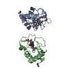
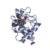
Mass: 618.503 Da / Num. of mol.: 2 / Source method: obtained synthetically / Formula: C34H34FeN4O4
#3: Water ChemComp-HOH / water


Mass: 18.015 Da / Num. of mol.: 120 / Source method: isolated from a natural source / Formula: H2O
Has protein modificationY
Nonpolymer detailsCOMPND MOLECULE: CYTOCHROME C. THE HEME IS OXIDIZED (FE 3+).

-
Experimental details

-
Experiment

ExperimentMethod: X-RAY DIFFRACTION

-
Sample preparation

CrystalDensity Matthews: 2.18 Å3/Da / Density % sol: 43.6 %
Crystal
*PLUS
Density % sol: 42 %
Crystal grow
*PLUS
pH: 7 / Method: vapor diffusion, hanging drop
Components of the solutions
*PLUS
IDConc.Common nameCrystal-IDSol-ID
130 mg/mlprotein1drop
220-25 %(w/v)PEG10001drop
30.05 Mpotassium phosphate1drop
430 %(w/v)PEG10001reservoir
50.05 Mpotassium phosphate1reservoir

-
Data collection

Diffraction sourceWavelength: 1.5418
DetectorType: XENTRONICS / Detector: AREA DETECTOR / Date: Jul 1, 1992
RadiationMonochromatic (M) / Laue (L): M / Scattering type: x-ray
Radiation wavelengthWavelength: 1.5418 Å / Relative weight: 1
ReflectionNum. obs: 8803 / % possible obs: 65 % / Redundancy: 4 % / Rmerge(I) obs: 0.0396
Reflection
*PLUS
Rmerge(I) obs: 0.0396

-
Processing

Software
NameClassification
PROFFTrefinement
PROLSQrefinement
XENGENdata reduction
RefinementResolution: 2.08→10 Å / σ(F): 2
Details: THE SIDE CHAINS OF RESIDUES LYS 72 AND LYS 87 IN MOLECULE A COULD BE FITTED TO THE ELECTRON DENSITY IN MORE THAN ONE CONFORMATION BUT THESE ARE NOT CLEARLY EXPRESSED ROTAMERS. THEY ARE ...Details: THE SIDE CHAINS OF RESIDUES LYS 72 AND LYS 87 IN MOLECULE A COULD BE FITTED TO THE ELECTRON DENSITY IN MORE THAN ONE CONFORMATION BUT THESE ARE NOT CLEARLY EXPRESSED ROTAMERS. THEY ARE IDENTIFIED AS 'C' IN THE ALTERNATE LOCATIONS COLUMN. THE SIDE CHAINS OF RESIDUES LYS 8, LYS 53, GLU 61, LYS 86 AND LYS 100 IN MOLECULE B HAVE ROTAMERS. THEY ARE DESIGNATED 'D' IN THE ALTERNATE CONFORMATIONS COLUMN. THE SIDE CHAINS OF RESIDUES LYS 22, LYS 72 AND LYS 88 IN MOLECULE B COULD BE FITTED TO THE ELECTRON DENSITY IN MORE THAN ONE CONFORMATION BUT THESE ARE NOT CLEARLY EXPRESSED ROTAMERS. THEY ARE IDENTIFIED AS 'D' IN THE ALTERNATE LOCATIONS COLUMN.
RfactorNum. reflection% reflection
obs0.177 7684 60 %
Displacement parametersBiso mean: 22.24 Å2
Refinement stepCycle: LAST / Resolution: 2.08→10 Å
ProteinNucleic acidLigandSolventTotal
Num. atoms1727 0 86 121 1934
Refine LS restraints
Refine-IDTypeDev idealDev ideal target
X-RAY DIFFRACTIONp_bond_d0.0170.02
X-RAY DIFFRACTIONp_angle_d0.0570.04
X-RAY DIFFRACTIONp_angle_deg
X-RAY DIFFRACTIONp_planar_d0.060.05
X-RAY DIFFRACTIONp_hb_or_metal_coord
X-RAY DIFFRACTIONp_mcbond_it0.081
X-RAY DIFFRACTIONp_mcangle_it0.11.5
X-RAY DIFFRACTIONp_scbond_it0.071
X-RAY DIFFRACTIONp_scangle_it0.10.15
X-RAY DIFFRACTIONp_plane_restr0.0120.02
X-RAY DIFFRACTIONp_chiral_restr0.1820.15
X-RAY DIFFRACTIONp_singtor_nbd0.2440.5
X-RAY DIFFRACTIONp_multtor_nbd0.3540.5
X-RAY DIFFRACTIONp_xhyhbond_nbd0.3230.5
X-RAY DIFFRACTIONp_xyhbond_nbd
X-RAY DIFFRACTIONp_planar_tor23
X-RAY DIFFRACTIONp_staggered_tor2215
X-RAY DIFFRACTIONp_orthonormal_tor2020
X-RAY DIFFRACTIONp_transverse_tor
X-RAY DIFFRACTIONp_special_tor
Software
*PLUS
Name: PROLSQ/PROFFT / Classification: refinement
LS refinement shell
*PLUS
Highest resolution: 2.08 Å / Lowest resolution: 2.35 Å / Total num. of bins used: 7 / Num. reflection obs: 1094 / Rfactor obs: 0.266

+
About Yorodumi

-
News

-
Feb 9, 2022. New format data for meta-information of EMDB entries

New format data for meta-information of EMDB entries

  • Version 3 of the EMDB header file is now the official format.
  • The previous official version 1.9 will be removed from the archive.

Related info.:EMDB header

External links:wwPDB to switch to version 3 of the EMDB data model

-
Aug 12, 2020. Covid-19 info

Covid-19 info

URL: https://pdbj.org/emnavi/covid19.php

New page: Covid-19 featured information page in EM Navigator.

Related info.:Covid-19 info / Mar 5, 2020. Novel coronavirus structure data

+
Mar 5, 2020. Novel coronavirus structure data

Novel coronavirus structure data

Related info.:Yorodumi Speices / Aug 12, 2020. Covid-19 info

External links:COVID-19 featured content - PDBj / Molecule of the Month (242):Coronavirus Proteases

+
Jan 31, 2019. EMDB accession codes are about to change! (news from PDBe EMDB page)

EMDB accession codes are about to change! (news from PDBe EMDB page)

  • The allocation of 4 digits for EMDB accession codes will soon come to an end. Whilst these codes will remain in use, new EMDB accession codes will include an additional digit and will expand incrementally as the available range of codes is exhausted. The current 4-digit format prefixed with “EMD-” (i.e. EMD-XXXX) will advance to a 5-digit format (i.e. EMD-XXXXX), and so on. It is currently estimated that the 4-digit codes will be depleted around Spring 2019, at which point the 5-digit format will come into force.
  • The EM Navigator/Yorodumi systems omit the EMD- prefix.

Related info.:Q: What is EMD? / ID/Accession-code notation in Yorodumi/EM Navigator

External links:EMDB Accession Codes are Changing Soon! / Contact to PDBj

+
Jul 12, 2017. Major update of PDB

Major update of PDB

  • wwPDB released updated PDB data conforming to the new PDBx/mmCIF dictionary.
  • This is a major update changing the version number from 4 to 5, and with Remediation, in which all the entries are updated.
  • In this update, many items about electron microscopy experimental information are reorganized (e.g. em_software).
  • Now, EM Navigator and Yorodumi are based on the updated data.

External links:wwPDB Remediation / Enriched Model Files Conforming to OneDep Data Standards Now Available in the PDB FTP Archive

-
Yorodumi

Thousand views of thousand structures

  • Yorodumi is a browser for structure data from EMDB, PDB, SASBDB, etc.
  • This page is also the successor to EM Navigator detail page, and also detail information page/front-end page for Omokage search.
  • The word "yorodu" (or yorozu) is an old Japanese word meaning "ten thousand". "mi" (miru) is to see.

Related info.:EMDB / PDB / SASBDB / Comparison of 3 databanks / Yorodumi Search / Aug 31, 2016. New EM Navigator & Yorodumi / Yorodumi Papers / Jmol/JSmol / Function and homology information / Changes in new EM Navigator and Yorodumi

Read more