 Movie
Movie Controller


+ Open data

- Basic information
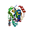
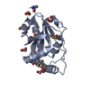
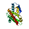
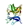
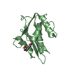
| Entry | Database: PDB / ID: 1cno | ||||||
|---|---|---|---|---|---|---|---|
| Title | STRUCTURE OF PSEUDOMONAS NAUTICA CYTOCHROME C552, BY MAD METHOD | ||||||
 Components Components | CYTOCHROME C552 | ||||||
 Keywords Keywords | ELECTRON TRANSPORT / PSEUDOMONAS NAUTICA / X RAY STRUCTURE / MULTIWAVELENGTH ANOMALOUS DISPERSION / HEME / CYTOCHROME C | ||||||
| Function / homology |  Function and homology information Function and homology informationperiplasmic space / electron transfer activity / heme binding / metal ion binding Similarity search - Function | ||||||
| Biological species |  Marinobacter hydrocarbonoclasticus (bacteria) Marinobacter hydrocarbonoclasticus (bacteria) | ||||||
| Method |  X-RAY DIFFRACTION / SYNCHROTRON / Resolution: 2.2 Å X-RAY DIFFRACTION / SYNCHROTRON / Resolution: 2.2 Å | ||||||
 Authors Authors | Brown, K. / Nurizzo, D. / Cambillau, C. | ||||||
 Citation Citation | Journal: J.Mol.Biol. / Year: 1999 Title: MAD structure of Pseudomonas nautica dimeric cytochrome c552 mimicks the c4 Dihemic cytochrome domain association. Authors: Brown, K. / Nurizzo, D. / Besson, S. / Shepard, W. / Moura, J. / Moura, I. / Tegoni, M. / Cambillau, C. | ||||||
| History |
|
- Structure visualization
| Structure viewer | Molecule: MolmilJmol/JSmol |
|---|
- Downloads & links
-Download
| PDBx/mmCIF format | 1cno.cif.gz | 156.4 KB | Display | PDBx/mmCIF format |
|---|---|---|---|---|
| PDB format | pdb1cno.ent.gz | 125.6 KB | Display | PDB format |
| PDBx/mmJSON format | 1cno.json.gz | Tree view | PDBx/mmJSON format | |
| Others |  Other downloads Other downloads |
-Validation report
| Arichive directory | https://data.pdbj.org/pub/pdb/validation_reports/cn/1cnoftp://data.pdbj.org/pub/pdb/validation_reports/cn/1cno | HTTPS FTP |
|---|
-Related structure data
| Similar structure data |
|---|


-Links
 PDBj
PDBj


- Assembly
| Deposited unit | 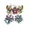
| ||||||||||||||||
|---|---|---|---|---|---|---|---|---|---|---|---|---|---|---|---|---|---|
| 1 | 
| ||||||||||||||||
| 2 | 
| ||||||||||||||||
| 3 | 
| ||||||||||||||||
| 4 | 
| ||||||||||||||||
| Unit cell |
| ||||||||||||||||
| Components on special symmetry positions |
| ||||||||||||||||
| Noncrystallographic symmetry (NCS) | NCS oper:
|
-Components
| #1: Protein | Mass: 8710.734 Da / Num. of mol.: 8 / Source method: isolated from a natural source Details: C HEME IN EACH MONOMER LINKED BY CYS 14 AND 17 AND HIS 18 AND MET 60 AS AXIAL LIGANDS Source: (natural) Marinobacter hydrocarbonoclasticus (bacteria)Cellular location: PERIPLASM / Strain: 617 / References: UniProt: P82903 #2: Chemical | ChemComp-HEC /   Mass: 618.503 Da / Num. of mol.: 8 / Source method: obtained synthetically / Formula: C34H34FeN4O4 Mass: 618.503 Da / Num. of mol.: 8 / Source method: obtained synthetically / Formula: C34H34FeN4O4#3: Chemical |   Mass: 92.094 Da / Num. of mol.: 3 / Source method: obtained synthetically / Formula: C3H8O3 Mass: 92.094 Da / Num. of mol.: 3 / Source method: obtained synthetically / Formula: C3H8O3#4: Water | ChemComp-HOH / |  Mass: 18.015 Da / Num. of mol.: 728 / Source method: isolated from a natural source / Formula: H2O Mass: 18.015 Da / Num. of mol.: 728 / Source method: isolated from a natural source / Formula: H2OHas protein modification | Y | Sequence details | THE 3 C-TERMINAL SIDE CHAINS WERE NOT SEEN IN THE DENSITY MAP AND WERE REPLACED BY ALANINE. LEU 36 ...THE 3 C-TERMINAL SIDE CHAINS WERE NOT SEEN IN THE DENSITY MAP AND WERE REPLACED BY ALANINE. LEU 36 CHANGED TO LYS 36; GLY 79 CHANGED TO TYR 79 COMPARED TO REF. EURO. J. BIOL. (1994), 224, 1011-1017. | |
|---|
-Experimental details
-Experiment
| Experiment | Method: X-RAY DIFFRACTION / Number of used crystals: 1 |
|---|
- Sample preparation
| Crystal | Density Matthews: 4.36 Å3/Da / Density % sol: 72 % Description: THE STARTING MODEL WAS THE C552 FROM PS. NAUTICA SOLVED BY MAD METHOD AT 2.8A RESOLUTION | ||||||||||||||||||||||||
|---|---|---|---|---|---|---|---|---|---|---|---|---|---|---|---|---|---|---|---|---|---|---|---|---|---|
| Crystal grow | pH: 5.6 / Details: pH 5.6 | ||||||||||||||||||||||||
| Crystal grow | *PLUS Temperature: 20 ℃ / Method: vapor diffusion | ||||||||||||||||||||||||
| Components of the solutions | *PLUS
|
-Data collection
| Diffraction | Mean temperature: 100 K |
|---|---|
| Diffraction source | Source: SYNCHROTRON / Site: ESRF  / Beamline: ID09 / Wavelength: 0.756 / Beamline: ID09 / Wavelength: 0.756 |
| Detector | Type: THOMPSON Detector: X RAY IMAGE INTENSIFIER (THOMPSON) + PRINCETON CCD DETECTOR Date: May 1, 1998 / Details: FOCUSED BEAM TOROIDAL |
| Radiation | Monochromator: LAUE BRAGG / Monochromatic (M) / Laue (L): M / Scattering type: x-ray |
| Radiation wavelength | Wavelength: 0.756 Å / Relative weight: 1 |
| Reflection | Resolution: 2.2→15 Å / Num. obs: 58183 / % possible obs: 99 % / Observed criterion σ(I): 2 / Redundancy: 3.5 % / Biso Wilson estimate: 18.84 Å2 / Rsym value: 0.108 / Net I/σ(I): 6.3 |
| Reflection shell | Resolution: 2.2→2.26 Å / Redundancy: 2.2 % / Mean I/σ(I) obs: 2 / Rsym value: 0.246 / % possible all: 94.3 |
| Reflection | *PLUS Rmerge(I) obs: 0.094 |
| Reflection shell | *PLUS Rmerge(I) obs: 0.229 |
- Processing
| Software |
| ||||||||||||||||||||||||||||||||||||||||||||||||||||||||||||
|---|---|---|---|---|---|---|---|---|---|---|---|---|---|---|---|---|---|---|---|---|---|---|---|---|---|---|---|---|---|---|---|---|---|---|---|---|---|---|---|---|---|---|---|---|---|---|---|---|---|---|---|---|---|---|---|---|---|---|---|---|---|
| Refinement | Resolution: 2.2→15 Å / Rfactor Rfree error: 0.005 / Data cutoff high absF: 400000 / Data cutoff low absF: 1.0E-5 / Cross valid method: THROUGHOUT / σ(F): 1
| ||||||||||||||||||||||||||||||||||||||||||||||||||||||||||||
| Displacement parameters | Biso mean: 19.26 Å2 | ||||||||||||||||||||||||||||||||||||||||||||||||||||||||||||
| Refine analyze | Luzzati d res low obs: 2.2 Å / Luzzati sigma a obs: 0.13 Å | ||||||||||||||||||||||||||||||||||||||||||||||||||||||||||||
| Refinement step | Cycle: LAST / Resolution: 2.2→15 Å
| ||||||||||||||||||||||||||||||||||||||||||||||||||||||||||||
| Refine LS restraints |
| ||||||||||||||||||||||||||||||||||||||||||||||||||||||||||||
| LS refinement shell | Resolution: 2.2→2.3 Å / Rfactor Rfree error: 0.016 / Total num. of bins used: 8
| ||||||||||||||||||||||||||||||||||||||||||||||||||||||||||||
| Software | *PLUS Name: X-PLOR / Version: 3.843 / Classification: refinement | ||||||||||||||||||||||||||||||||||||||||||||||||||||||||||||
| Refinement | *PLUS Rfactor Rfree: 0.2083 | ||||||||||||||||||||||||||||||||||||||||||||||||||||||||||||
| Solvent computation | *PLUS | ||||||||||||||||||||||||||||||||||||||||||||||||||||||||||||
| Displacement parameters | *PLUS Biso mean: 19.267 Å2 | ||||||||||||||||||||||||||||||||||||||||||||||||||||||||||||
| Refine LS restraints | *PLUS
|
