 Movie
Movie Controller

[English] 日本語
 Yorodumi
- PDB-1cjf: PROFILIN BINDS PROLINE-RICH LIGANDS IN TWO DISTINCT AMIDE BACKBON... -
+ Open data

- Basic information
| Entry | Database: PDB / ID: 1cjf | ||||||
|---|---|---|---|---|---|---|---|
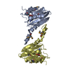
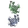
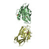
| Title | PROFILIN BINDS PROLINE-RICH LIGANDS IN TWO DISTINCT AMIDE BACKBONE ORIENTATIONS | ||||||
 Components Components |
| ||||||
 Keywords Keywords | STRUCTURAL REGULATION PROTEIN / PROFILIN / ACTIN-BINDING PROTEIN / CYTOSKELETON / POLY-L-PROLINE | ||||||
| Function / homology |  Function and homology information Function and homology informationsynapse maturation / adenyl-nucleotide exchange factor activity / negative regulation of actin filament bundle assembly / modification of postsynaptic actin cytoskeleton / positive regulation of actin filament bundle assembly / regulation of actin filament polymerization / Signaling by ROBO receptors / negative regulation of stress fiber assembly / proline-rich region binding / PCP/CE pathway ...synapse maturation / adenyl-nucleotide exchange factor activity / negative regulation of actin filament bundle assembly / modification of postsynaptic actin cytoskeleton / positive regulation of actin filament bundle assembly / regulation of actin filament polymerization / Signaling by ROBO receptors / negative regulation of stress fiber assembly / proline-rich region binding / PCP/CE pathway / positive regulation of actin filament polymerization / positive regulation of ruffle assembly / positive regulation of epithelial cell migration / actin monomer binding / phosphotyrosine residue binding / phosphatidylinositol-4,5-bisphosphate binding / RHO GTPases Activate Formins / modulation of chemical synaptic transmission / small GTPase binding / Platelet degranulation / actin cytoskeleton organization / actin binding / blood microparticle / cell cortex / cytoskeleton / protein stabilization / cadherin binding / focal adhesion / glutamatergic synapse / RNA binding / extracellular exosome / membrane / nucleus / cytosol / cytoplasm Similarity search - Function | ||||||
| Biological species |  Homo sapiens (human) Homo sapiens (human) | ||||||
| Method |  X-RAY DIFFRACTION / MOLECULAR REPLACEMENT / Resolution: 2.3 Å X-RAY DIFFRACTION / MOLECULAR REPLACEMENT / Resolution: 2.3 Å | ||||||
 Authors Authors | Mahoney, N.M. / Fedorov, A.A. / Fedorov, E. / Rozwarski, D.A. / Almo, S.C. | ||||||
 Citation Citation | Journal: Nat.Struct.Biol. / Year: 1999 Title: Profilin binds proline-rich ligands in two distinct amide backbone orientations. Authors: Mahoney, N.M. / Rozwarski, D.A. / Fedorov, E. / Fedorov, A.A. / Almo, S.C. | ||||||
| History |
|
- Structure visualization
| Structure viewer | Molecule: MolmilJmol/JSmol |
|---|
- Downloads & links
-Download
| PDBx/mmCIF format | 1cjf.cif.gz | 69.3 KB | Display | PDBx/mmCIF format |
|---|---|---|---|---|
| PDB format | pdb1cjf.ent.gz | 51.7 KB | Display | PDB format |
| PDBx/mmJSON format | 1cjf.json.gz | Tree view | PDBx/mmJSON format | |
| Others |  Other downloads Other downloads |
-Validation report
| Arichive directory | https://data.pdbj.org/pub/pdb/validation_reports/cj/1cjfftp://data.pdbj.org/pub/pdb/validation_reports/cj/1cjf | HTTPS FTP |
|---|
-Related structure data
| Related structure data | 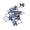 1cf0C 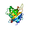 1filS S: Starting model for refinement C: citing same article ( |
|---|---|
| Similar structure data |


-Links
 PDBj
PDBj


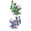
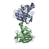
- Assembly
| Deposited unit | 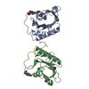
| ||||||||
|---|---|---|---|---|---|---|---|---|---|
| 1 |
| ||||||||
| Unit cell |
| ||||||||
| Noncrystallographic symmetry (NCS) | NCS oper: (Code: given Matrix: (-0.066716, -0.003106, 0.997767), Vector: |
-Components
| #1: Protein | Mass: 14940.021 Da / Num. of mol.: 2 Source method: isolated from a genetically manipulated source Details: HMC FLUOROPHORE / Source: (gene. exp.) Homo sapiens (human) / Strain: BL21(DE3) / Cellular location: CYTOPLASM / References: UniProt: P07737#2: Protein/peptide | Mass: 1474.737 Da / Num. of mol.: 2 / Source method: obtained synthetically #3: Chemical |   Mass: 220.221 Da / Num. of mol.: 2 / Source method: obtained synthetically / Formula: C12H12O4 Mass: 220.221 Da / Num. of mol.: 2 / Source method: obtained synthetically / Formula: C12H12O4#4: Water | ChemComp-HOH / |  Mass: 18.015 Da / Num. of mol.: 24 / Source method: isolated from a natural source / Formula: H2O Mass: 18.015 Da / Num. of mol.: 24 / Source method: isolated from a natural source / Formula: H2O |
|---|
-Experimental details
-Experiment
| Experiment | Method: X-RAY DIFFRACTION / Number of used crystals: 1 |
|---|
- Sample preparation
| Crystal | Density Matthews: 2.6 Å3/Da / Density % sol: 53 % | ||||||||||||||||||||||||||||||||||||||||||
|---|---|---|---|---|---|---|---|---|---|---|---|---|---|---|---|---|---|---|---|---|---|---|---|---|---|---|---|---|---|---|---|---|---|---|---|---|---|---|---|---|---|---|---|
| Crystal grow | pH: 7.5 Details: 1.4 M AMMONIUM SULFATE, 0.1 M HEPES PH 7.5, 0.1 M NACL | ||||||||||||||||||||||||||||||||||||||||||
| Crystal grow | *PLUS Method: vapor diffusion, hanging dropDetails: drop consists of equal volume of protein and reservoir solutions | ||||||||||||||||||||||||||||||||||||||||||
| Components of the solutions | *PLUS
|
-Data collection
| Diffraction | Mean temperature: 298 K |
|---|---|
| Diffraction source | Source: ROTATING ANODE / Type: RIGAKU RU200 / Wavelength: 1.5418 |
| Detector | Type: SIEMENS X1000 / Detector: AREA DETECTOR / Date: Jul 1, 1998 / Details: MIRRORS |
| Radiation | Monochromator: GRAPHITE CRYSTAL / Protocol: SINGLE WAVELENGTH / Monochromatic (M) / Laue (L): M / Scattering type: x-ray |
| Radiation wavelength | Wavelength: 1.5418 Å / Relative weight: 1 |
| Reflection | Resolution: 2.3→8 Å / Num. obs: 11957 / % possible obs: 90 % / Observed criterion σ(I): 2 / Rmerge(I) obs: 0.052 |
- Processing
| Software |
| ||||||||||||||||||||||||||||||||||||||||||||||||||||||||||||
|---|---|---|---|---|---|---|---|---|---|---|---|---|---|---|---|---|---|---|---|---|---|---|---|---|---|---|---|---|---|---|---|---|---|---|---|---|---|---|---|---|---|---|---|---|---|---|---|---|---|---|---|---|---|---|---|---|---|---|---|---|---|
| Refinement | Method to determine structure: MOLECULAR REPLACEMENT Starting model: 1FIL Resolution: 2.3→8 Å / Data cutoff high absF: 1000000 / Data cutoff low absF: 0.001 / Cross valid method: THROUGHOUT / σ(F): 2
| ||||||||||||||||||||||||||||||||||||||||||||||||||||||||||||
| Displacement parameters | Biso mean: 22.15 Å2 | ||||||||||||||||||||||||||||||||||||||||||||||||||||||||||||
| Refine analyze |
| ||||||||||||||||||||||||||||||||||||||||||||||||||||||||||||
| Refinement step | Cycle: LAST / Resolution: 2.3→8 Å
| ||||||||||||||||||||||||||||||||||||||||||||||||||||||||||||
| Refine LS restraints |
| ||||||||||||||||||||||||||||||||||||||||||||||||||||||||||||
| Software | *PLUS Name: X-PLOR / Version: 3.851 / Classification: refinement | ||||||||||||||||||||||||||||||||||||||||||||||||||||||||||||
| Refine LS restraints | *PLUS
|
